Two-Dimensional Image-Based Respiratory Navigator for Free-Breathing Coronary Magnetic Resonance Angiography
- Affiliations
-
- 1Division of Mechanical and Biomedical Engineering, Ewha Womans University, Seoul, Korea. taehoons@ewha.ac.kr
- KMID: 2408821
- DOI: http://doi.org/10.13104/imri.2018.22.1.71
Abstract
- PURPOSE
To develop a two-dimensional (2D) image-based respiratory motion correction technique for free-breathing coronary magnetic resonance angiography (MRA).
MATERIALS AND METHODS
The proposed respiratory navigator obtained aliased a 2D sagittal image from under-sampled k-space data and utilized motion correlation between the aliased images. The proposed navigator was incorporated into the conventional coronary MRA sequence including the diaphragm navigator and tested in three healthy subjects.
RESULTS
The delineation of major coronary arteries was significantly improved using the proposed 2D motion correction (S/I and A/P) compared to one-dimensional (S/I) correction using the conventional diaphragm navigator.
CONCLUSION
The 2D image-based respiratory navigator was proposed for free-breathing coronary angiography and showed the potential for improving respiratory motion correction compared to the conventional 1D correction.
MeSH Terms
Figure
-
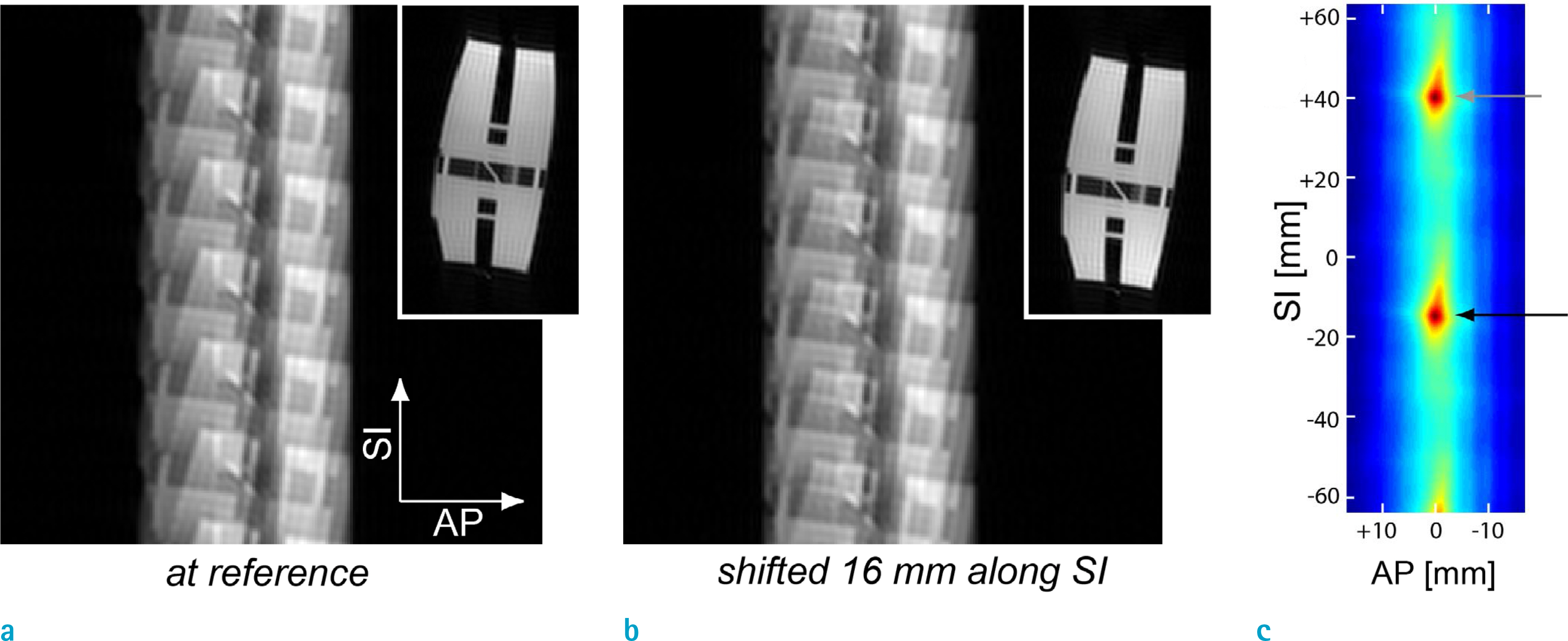
Fig. 1. Illustration of the proposed 2D motion estimation. (a) Six-fold aliased image of the resolution phantom (a): at a reference position, (b) Shifted 16 mm along the SI direction. (c) Correlation between the reference aliased image and the second aliased image shifted over 2D search space. Due to the same translations of the six replicas, a local maximum is yielded at the true displacement (black arrow), and also at increments of (n·FOV/R) (gray arrow) away from the true solution (n = integer, R = under-sampling rate).

Fig. 2. Timing diagram of coronary MRA with diaphragm navigator and 2D image-based navigator. The sequence consisted of a T2-preparation pulse, diaphragm navigator acquisition, fat saturation pulse, coronary acquisition, notched saturation pulse, and 2D image-based navigator acquisition, sequentially.
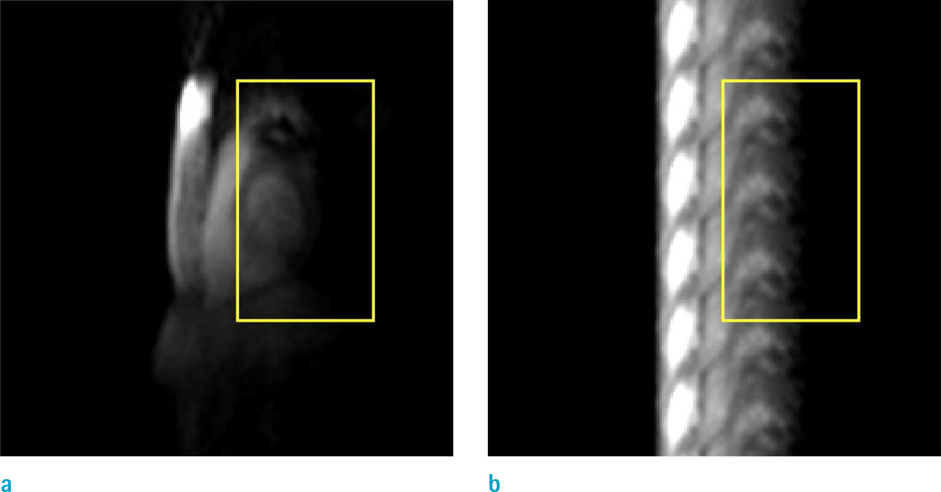
Fig. 3. (a) Full-FOV sagittal image was acquired prior to coronary MRA scan and used to specify an ROI for motion estimation (yellow box). (b) Six-fold aliased image acquired during an MRA scan clearly shows image features to be used for motion estimation.
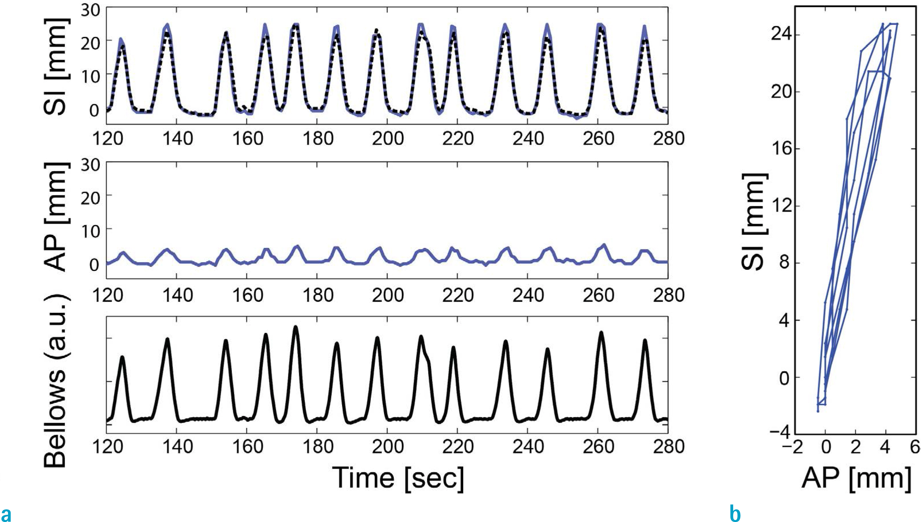
Fig. 4. (a) Estimated motion in the S/I and A/P directions (in millimeters) and bellows signal (in arbitrary unit denoted as ‘a.u.’). In the top row, blue solid curve and black dotted curve represents the estimates by the proposed image-based navigator and the diaphragm navigator, respectively. (b) Trajectory of estimated motion in the SI-AP plane.

Fig. 5. Representative coronary MRA images obtained using no correction (left column), 1D correction (S/I) based on the diaphragm navigator (middle row) and 2D correction (S/I + A/P) based on the proposed image-based navigator (right column). The delineation of the arterial tree progressively improves with the 1D and 2D corrections (arrows).
Reference
-
References
1. Stuber M, Weiss RG. Coronary magnetic resonance angiography. J Magn Reson Imaging. 2007; 26:219–234.
Article2. Chiribiri A, Ishida M, Nagel E, Botnar RM. Coronary imaging with cardiovascular magnetic resonance: current state of the art. Prog Cardiovasc Dis. 2011; 54:240–252.
Article3. Stehning C, Bornert P, Nehrke K, Eggers H, Stuber M. Free-breathing whole-heart coronary MRA with 3D radial SSFP and self-navigated image reconstruction. Magn Reson Med. 2005; 54:476–480.
Article4. Danias PG, McConnell MV, Khasgiwala VC, Chuang ML, Edelman RR, Manning WJ. Prospective navigator correction of image position for coronary MR angiography. Radiology. 1997; 203:733–736.
Article5. Danias PG, Stuber M, Botnar RM, Kissinger KV, Edelman RR, Manning WJ. Relationship between motion of coronary arteries and diaphragm during free breathing: lessons from realtime MR imaging. AJR Am J Roentgenol. 1999; 172:1061–1065.
Article6. Nehrke K, Bornert P, Manke D, Bock JC. Free-breathing cardiac MR imaging: study of implications of respiratory motion–initial results. Radiology. 2001; 220:810–815.
Article7. Taylor AM, Keegan J, Jhooti P, Firmin DN, Pennell DJ. Calculation of a subject-specific adaptive motion-correction factor for improved realtime navigator echo-gated magnetic resonance coronary angiography. J Cardiovasc Magn Reson. 1999; 1:131–138.
Article8. Keegan J, Gatehouse P, Yang GZ, Firmin D. Coronary artery motion with the respiratory cycle during breathholding and free-breathing: implications for slice-followed coronary artery imaging. Magn Reson Med. 2002; 47:476–481.
Article9. Lai P, Larson AC, Bi X, Jerecic R, Li D. A dual-projection respiratory self-gating technique for whole-heart coronary MRA. J Magn Reson Imaging. 2008; 28:612–620.
Article10. Lai P, Bi X, Jerecic R, Li D. A respiratory self-gating technique with 3D-translation compensation for free-breathing whole-heart coronary MRA. Magn Reson Med. 2009; 62:731–738.
Article11. Brittain JH, Hu BS, Wright GA, Meyer CH, Macovski A, Nishimura DG. Coronary angiography with magnetization-prepared T2 contrast. Magn Reson Med. 1995; 33:689–696.12. Cunningham CH, Lustig M, Hu BS, et al. Novel design for notched RF saturation pulses using the SLR transform. In Proceedings of the 15th Scientific Meeting of International Society for Magnetic Resonance in Medicine. Berlin. 2007. 1709.13. Manke D, Nehrke K, Bornert P, Rosch P, Dossel O. Respiratory motion in coronary magnetic resonance angiography: a comparison of different motion models. J Magn Reson Imaging. 2002; 15:661–671.
Article
- Full Text Links
-
- Actions
-
Cited
- CITED
-
- Close
- Share
-
- Similar articles
-
- Cardiac MR Assessment of Coronary Arteries
- Feasibility and Diagnostic Accuracy of Whole Heart Coronary MR Angiography Using Free-Breathing 3D Balanced Turbo-Field-Echo with SENSE and the Half-Fourier Acquisition Technique
- Two-Dimensional Breath-Hold Coronary MR Angiography in Normal Adults
- Clinical Utility of Magnetic Resonance Angiography
- Myocardial Viability: Comparison of Free-Breathing Navigator-echo-gated Three-Dimensional Inversion-Recovery Gradient-Echo MR and Standard Multiple Breath-Hold Two-Dimensional Inversion-Recovery Gradient-Echo MR
