Lymphoepithelial Cyst in Pancreas: a Case Report with Magnetic Resonance Imaging Findings
- Affiliations
-
- 1Department of Radiology, Dankook University Hospital, Chungnam, Korea. deepva@hanmail.net
- KMID: 2408819
- DOI: http://doi.org/10.13104/imri.2018.22.1.61
Abstract
- Pancreatic lymphoepithelial cysts (LECs) are rare pancreatic cysts with squamous epithelial lining surrounded by dense lymphoid tissue. A preoperative diagnosis of LECs is difficult due to imaging diversity and inadequate documentation because of their rarity. We present a case of surgically confirmed pancreatic LEC with magnetic resonance imaging (MRI) findings as heterogeneous signal intensity on T2-weighted images with multiple septa-like structures, slightly hypo-signal intensity on T1-weighted images, and thin-wall enhancement on dynamic contrast images. LECs are benign lesions without any malignant potential. Therefore, the inclusion of LEC in the differential diagnosis of cystic pancreatic lesions may reduce unnecessary surgical procedures.
MeSH Terms
Figure
-
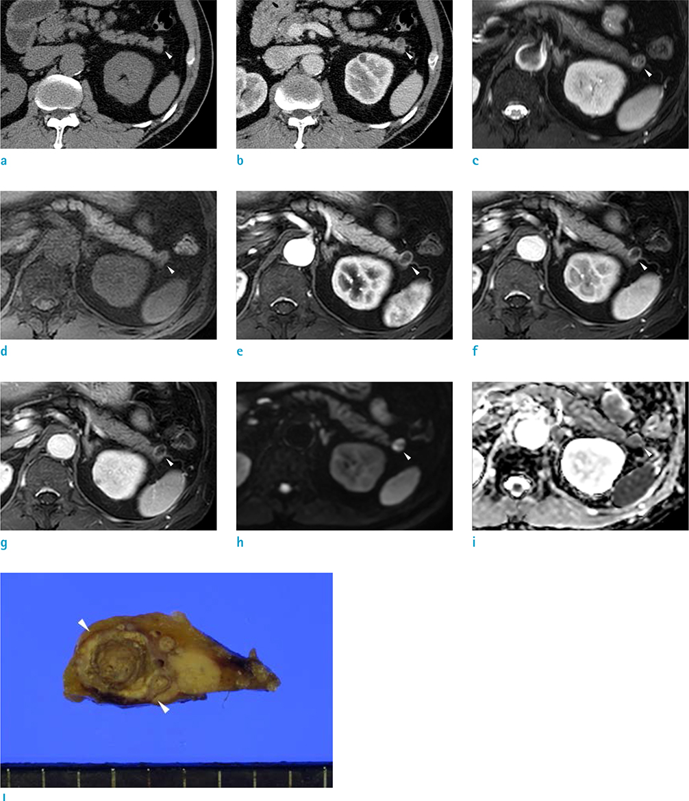
Fig. 1 A lymphoepithelial cyst of the pancreas in a 59-year-old man. A 1.3-cm-sized well-defined oval lesion is seen in the pancreatic tail portion with an exophytic appearance. The lesion shows isoattenuation with pancreas on precontrast CT scan (a) and thin peripheral wall enhancement on postcontrast CT scan (b). On T2-weighted image (c), the lesion shows heterogeneous signal intensity with multiple septa-like structures present inside. The lesion shows slightly hypo-signal intensity on the precontrast T1-weighted image (d) with thin peripheral wall enhancement on arterial (e), portal (f), and 3-min delayed phase (g) images of a dynamic contrast enhancement study. The lesion shows high signal intensity on axial high b-value image (b = 800 s/mm2) (h); however, the ADC map (i) reveals no diffusion restriction. Photograph of the gross surgical specimen (j) demonstrates cystic nodule with the pale yellowish surface, filled with keratin material, and encapsulated by a fibrous capsule.
Reference
-
1. Brugge WR, Lauwers GY, Sahani D, Fernandez-del Castillo C, Warshaw AL. Cystic neoplasms of the pancreas. N Engl J Med. 2004; 351:1218–1226.
Article2. Kavuturu S, Sarwani NE, Ruggeiro FM, et al. Lymphoepithelial cysts of the pancreas. Can preoperative imaging distinguish this benign lesion from malignant or pre-malignant cystic pancreatic lesions? JOP. 2013; 14:250–225.3. Ramsden KL, Newman J. Lymphoepithelial cyst of the pancreas. Histopathology. 1991; 18:267–268.
Article4. Adsay NV, Hasteh F, Cheng JD, et al. Lymphoepithelial cysts of the pancreas: a report of 12 cases and a review of the literature. Mod Pathol. 2002; 15:492–501.
Article5. Nam SJ, Hwang HK, Kim H, et al. Lymphoepithelial cysts in the pancreas: MRI of two cases with emphasis of diffusion-weighted imaging characteristics. J Magn Reson Imaging. 2010; 32:692–696.
Article6. Kim YH, Auh YH, Kim KW, Lee MG, Kim KS, Park SY. Lymphoepithelial cysts of the pancreas: CT and sonographic findings. Abdom Imaging. 1998; 23:185–187.
Article7. Chen S, Ikawa F, Kurisu K, Arita K, Takaba J, Kanou Y. Quantitative MR evaluation of intracranial epidermoid tumors by fast fluid-attenuated inversion recovery imaging and echo-planar diffusion-weighted imaging. AJNR Am J Neuroradiol. 2001; 22:1089–1096.8. Fukukura Y, Inoue H, Miyazono N, et al. Lymphoepithelial cysts of the pancreas: demonstration of lipid component using CT and MRI. J Comput Assist Tomogr. 1998; 22:311–313.9. Ahlawat SK. Lymphoepithelial cyst of pancreas. Role of endoscopic ultrasound guided fine needle aspiration. JOP. 2008; 9:230–234.10. Centeno BA, Stockwell JW, Lewandrowski KB. Cyst fluid cytology and chemical features in a case of lymphoepithelial cyst of the pancreas: a rare and difficult preoperative diagnosis. Diagn Cytopathol. 1999; 21:328–330.
Article
- Full Text Links
-
- Actions
-
Cited
- CITED
-
- Close
- Share
-
- Similar articles
-
- Lymphoepithelial Cyst of the Pancreas: A Case Report
- Lymphoepithelial Cyst of the Pancreas: A Case Report
- A lymphoepithelial cyst of the pancreas: A case report
- A Case of Lymphoepithelial Cyst of the Pancreas Showing Characteristic Findings on Endoscopic Ultrasonography
- Squamous Cell Carcinoma of the Pancreas: A Case Report
