Gelatinous Transformation of Bone Marrow Mimicking Malignant Marrow-Replacing Lesion on Magnetic Resonance Imaging in a Patient without Underlying Devastating Disease
- Affiliations
-
- 1Department of Radiology, Gangnam Severance Hospital, Yonsei University College of Medicine, Seoul, Korea. agn70@yuhs.ac
- 2Department of Pathology, Green Cross Laboratories, Gyeonggi-do, Korea.
- 3Department of Orthopaedic Surgery, Gangnam Severance Hospital, Yonsei University College of Medicine, Seoul, Korea.
- KMID: 2408817
- DOI: http://doi.org/10.13104/imri.2018.22.1.50
Abstract
- Gelatinous transformation of bone marrow is characterized by hypoplasia of fat cells with focal loss of hematopoietic cells and deposition of extracellular gelatinous substances. It is known to be associated with devastating underlying diseases that starve bone marrow. Here, we present a case of a patient whose magnetic resonance (MR) imaging findings of vertebral column were interpreted as metastasis or hematologic malignancy, however, the final diagnosis revealed a gelatinous transformation of bone marrow. This is the first report of gelatinous transformation of bone marrow without evidence of underlying devastating disease.
Keyword
MeSH Terms
Figure
-
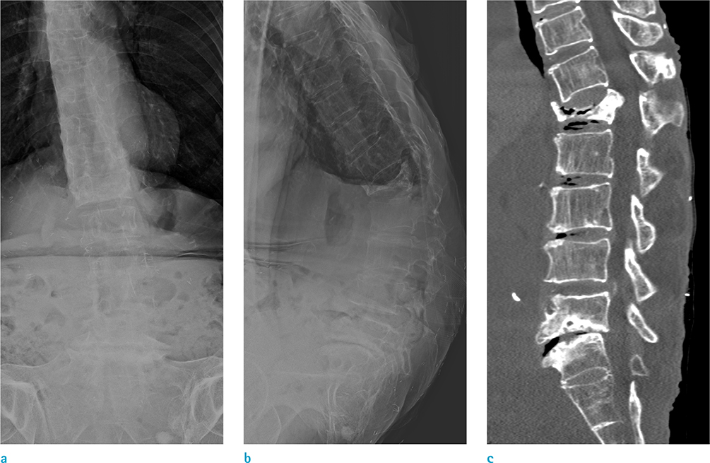
Fig. 1 Simple radiographs and computed tomography (CT) images of the lumbar spine obtained from a 75-year-old female who developed gelatinous transformation of the bone marrow. (a, b) Anterior and lateral lumbar spine radiographs revealed kyphosis and scoliosis of the thoracic-lumbar spine, with a wedging deformity of the L1 vertebral body. (c) CT shows a burst fracture of the L1 vertebral body without obvious osteolytic lesion. Diffuse lack of fat tissue on CT in and around the muscle is also seen.
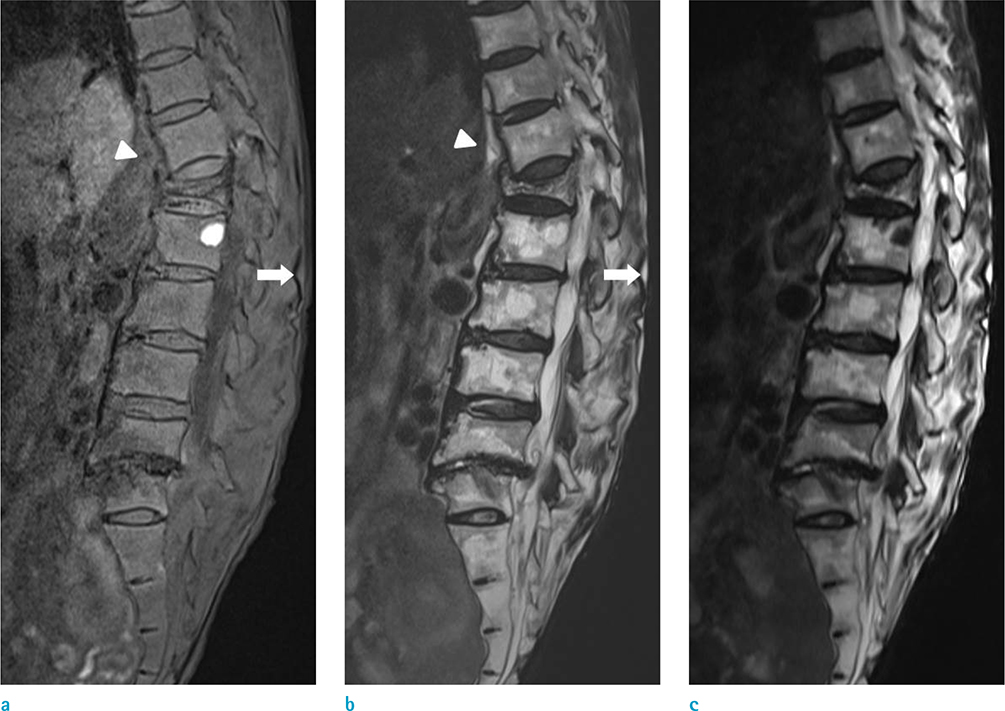
Fig. 2 Sagittal magnetic resonance (MR) imaging of the lumbar spine. (a) The spinal bone marrow shows extensive hypointense signal involving the entire vertebral column on a T1-weighted (repetition time [TR]: 450 ms, echo time [TE]: 9.8 ms) image. Diffuse hypointense signal intensity of retroperitoneal fat (arrowhead) and subcutaneous fat (arrow) on T1-weighted image is also seen. Extensive loss of fat in and around the muscles and in the subcutaneous layer at the back, suggests severe malnutrition. (b) T2-weighted MR (TR: 4192 ms, TE: 100 ms) image shows irregular and patchy hyperintense signals of vertebral marrow with respect to skeletal muscle. Hyperintense signal intensity of retroperitoneal fat (arrowhead) and subcutaneous fat (arrow) on T2-weighted image is also seen. (c) Sagittal fat-saturated T2-weighted (TR: 4580 ms, TE: 113 ms) images show hyperintense signals interspersed in L-spine, which indicates that the lesions are not fatty marrow.
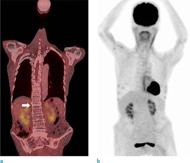
Fig. 3 18F-fluorodeoxyglucose (FDG) positron emission tomography (PET)-computed tomography (CT) of the whole body. (a, b) 18F-FDG-PET-CT scan revealed no hypermetabolic foci to suggest primary malignancy or distant metastasis, associated with the compressed L1 vertebral body (arrow).
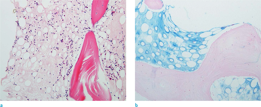
Fig. 4 A biopsy specimen of the bilateral posterior superior iliac crest shows bone marrow with gelatinous transformation. (a) Fat atrophy, hypoplasia of trilineage hematopoietic cells, and accumulation of amorphous extracellular material are seen (Hematoxylin & Eosin staining, × 200). (b) The extracellular gelatinous substance tested positive for Alcian blue stain (Alcian blue, × 100).
Reference
-
1. Bohm J. Gelatinous transformation of the bone marrow: the spectrum of underlying diseases. Am J Surg Pathol. 2000; 24:56–65.2. Sen R, Singh S, Singh H, Gupta A, Sen J. Clinical profile in gelatinous bone marrow transformation. J Assoc Physicians India. 2003; 51:585–588.3. Seaman JP, Kjeldsberg CR, Linker A. Gelatinous transformation of the bone marrow. Hum Pathol. 1978; 9:685–692.
Article4. Vande Berg BC, Malghem J, Devuyst O, Maldague BE, Lambert MJ. Anorexia nervosa: correlation between MR appearance of bone marrow and severity of disease. Radiology. 1994; 193:859–864.
Article5. Hanrahan CJ, Shah LM. MRI of spinal bone marrow: part 2, T1-weighted imaging-based differential diagnosis. AJR Am J Roentgenol. 2011; 197:1309–1321.
Article6. Feng CS. Gelatinous transformation of marrow in a case of acute myelogenous leukemia post-chemotherapy. Am J Hematol. 1991; 38:220–222.
Article7. Ifrah N, Saint-Andre JP, de Gentile L, et al. Gelatinous transformation of the bone marrow: manifestation of an acute leukemia. Acta Haematol. 1989; 82:165–168.
Article8. Chong A, Song HC, Oh JR, et al. Gelatinous degeneration of the bone marrow mimicking osseous metastasis on 18F-FDG PET/CT. Clin Nucl Med. 2012; 37:798–800.
Article9. Brennan CM, Atkins KA, Druzgal CH, Gaskin CM. Magnetic resonance imaging appearance of scurvy with gelatinous bone marrow transformation. Skeletal Radiol. 2012; 41:357–360.
Article10. Stroup JS, Stephens JR, Baker DL. Gelatinous bone marrow in an HIV-positive patient. Proc (Bayl Univ Med Cent). 2007; 20:254–256.
Article
- Full Text Links
-
- Actions
-
Cited
- CITED
-
- Close
- Share
-
- Similar articles
-
- A Case of Anorexia Nervosa with Gelatinous Transformation of Marrow
- A Case of Gelatinous Transformation of Marrow in a Patient with Congestive Heart Failure in Malnutrition
- MR Imaging of the Bone Marrow
- Differential Diagnosis of Vertebral Lesion by Magnetic Resonance Imaging
- Gelatinous transformation of the bone marrow in hepatocellular carcinoma
