Correlation of Semi-Quantitative Breast-Specific Gamma Imaging Findings with Dynamic Contrast-Enhanced MRI Parameters Assessed by a Computer-Aided Evaluation Program for Breast Cancer
- Affiliations
-
- 1Department of Radiology, Hanyang University College of Medicine, Seoul, Korea. huilingkoo@gmail.com
- 2Department of Radiology, Hanyang University College of Medicine, Hanyang University Guri Hospital, Guri, Korea.
- 3Department of Nuclear Medicine, Hanyang University College of Medicine, Seoul, Korea.
- 4Department of General Surgery, Hanyang University College of Medicine, Seoul, Korea.
- KMID: 2408258
- DOI: http://doi.org/10.3348/jksr.2018.78.2.95
Abstract
- PURPOSE
To investigate whether a correlation exists between the semi-quantitative breast-specific gamma imaging (BSGI) findings and dynamic contrast-enhanced (DCE) MRI parameters assessed by a computer-aided evaluation program.
MATERIALS AND METHODS
Semi-quantitative index of the lesion to non-lesion (L/N) ratio in BSGI and DCE-MRI parameters was assessed by a computer-aided evaluation program, where 47 cases of invasive breast cancer were obtained. Correlations between the L/N ratio and DCE-MRI parameters were assessed by a computer-aided evaluation program. Tumor diameter (cm), angio-volume (cc), degree of initial peak enhancement (%), persistent enhancement proportion (%), and washout enhancement proportion (%) were analysed. The relationships between the L/N ratio and DCE-MRI parameters were evaluated by a univariate and multivariate regression analysis.
RESULTS
The mean L/N ratio of the 47 tumors was 3.63 ± 2.19 (range: 1-13.1). The L/N ratio was higher in tumors with larger diameters (p < 0.001), increased angio-volume (p < 0.001), higher degree of initial peak enhancement (p = 0.005) and increased washout enhancement proportion (p = 0.004). In the multivariate regression analysis, angio-volume (cc) and washout enhancement proportion (%) were associated with L/N ratio (p = 0.007 and p = 0.024, respectively).
CONCLUSION
There was a correlation between the semi-quantitative L/N ratio in BSGI and DCE-MRI parameters assessed by a computer-aided evaluation program for breast cancer.
MeSH Terms
Figure
-
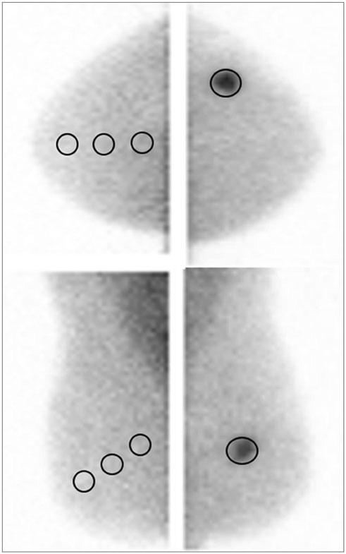
Fig. 1 Lesion to non-lesion ratio of breast cancer in the left upper outer breast. For lesions, the maximum ROI covering the lesion is drawn at the craniocaudal view and the mediolateral oblique view. For non-lesions, another three circular ROIs, approximately 1 cm in diameter, are drawn in the contralateral trisectioned breast, parallel to the base of the breast (anterior/middle/posterior). ROI = region of interest
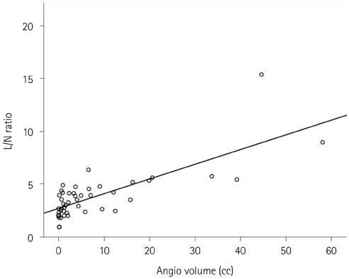
Fig. 2 Scatter plot of L/N ratio in breast-specific gamma imaging and angio-volume (coefficient of determination, R2 = 0.58). L/N = lesion to non-lesion
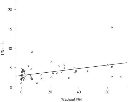
Fig. 3 Scatter plot of L/N ratio in breast-specific gamma imaging and washout enhancement proportion (coefficient of determination, R2 = 0.16). L/N = lesion to non-lesion

Fig. 4 Invasive ductal carcinoma in a 47 year-old woman. A. Rt. cranio-caudal view of breast-specific gamma imaging. Lesion to non-lesion ratio is 3.97. B. Angiomap and volume rendering image of the tumor obtained by computer-aided evaluation program in DCE-MRI (diameters: 2.9 × 2.0 × 2.2 cm, angio volume: 5.0 cc and washout enhancement proportion: 39%).
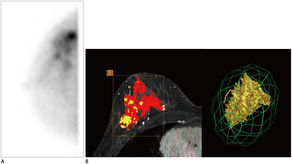
Fig. 5 Invasive ductal carcinoma in a 54 year-old woman. A. Rt. cranio-caudal view of breast-specific gamma imaging. Lesion to non-lesion ratio is 8.99. B. Angiomap and volume rendering image of the tumor obtained by computer-aided evaluation program in DCE-MRI (diameters: 6.9 × 5.1 × 8.1 cm, angio volume: 57.9 cc, washout enhancement proportion: 76%).
Reference
-
1. Brem RF, Rechtman LR. Nuclear medicine imaging of the breast: a novel, physiologic approach to breast cancer detection and diagnosis. Radiol Clin North Am. 2010; 48:1055–1074.
Article2. Yoon HJ, Kim Y, Chang KT, Kim BS. Prognostic value of semi-quantitative tumor uptake on Tc-99m sestamibi breast-specific gamma imaging in invasive ductal breast cancer. Ann Nucl Med. 2015; 29:553–560.
Article3. Yu X, Hu G, Zhang Z, Qiu F, Shao X, Wang X, et al. Retrospective and comparative analysis of (99m)Tc-Sestamibi breast specific gamma imaging versus mammography, ultrasound, and magnetic resonance imaging for the detection of breast cancer in Chinese women. BMC Cancer. 2016; 16:450.
Article4. Specht JM, Mankoff DA. Advances in molecular imaging for breast cancer detection and characterization. Breast Cancer Res. 2012; 14:206.
Article5. Sun Y, Wei W, Yang HW, Liu JL. Clinical usefulness of breast-specific gamma imaging as an adjunct modality to mammography for diagnosis of breast cancer: a systemic review and meta-analysis. Eur J Nucl Med Mol Imaging. 2013; 40:450–463.
Article6. Tan H, Jiang L, Gu Y, Xiu Y, Han L, Wu P, et al. Visual and semi-quantitative analyses of dual-phase breast-specific gamma imaging with Tc-99m-sestamibi in detecting primary breast cancer. Ann Nucl Med. 2014; 28:17–24.
Article7. Kuhl CK, Mielcareck P, Klaschik S, Leutner C, Wardelmann E, Gieseke J, et al. Dynamic breast MR imaging: are signal intensity time course data useful for differential diagnosis of enhancing lesions? Radiology. 1999; 211:101–110.
Article8. Orel SG, Schnall MD. MR imaging of the breast for the detection, diagnosis, and staging of breast cancer. Radiology. 2001; 220:13–30.
Article9. Jena A, Taneja S, Singh A, Negi P, Mehta SB, Sarin R. Role of pharmacokinetic parameters derived with high temporal resolution DCE MRI using simultaneous PET/MRI system in breast cancer: a feasibility study. Eur J Radiol. 2017; 86:261–266.
Article10. Aghaei F, Tan M, Hollingsworth AB, Qian W, Liu H, Zheng B. Computer-aided breast MR image feature analysis for prediction of tumor response to chemotherapy. Med Phys. 2015; 42:6520–6528.
Article11. Dorrius MD, Jansen-van der Weide MC, van Ooijen PM, Pijnappel RM, Oudkerk M. Computer-aided detection in breast MRI: a systematic review and meta-analysis. Eur Radiol. 2011; 21:1600–1608.
Article12. Kim BS. Usefulness of breast-specific gamma imaging as an adjunct modality in breast cancer patients with dense breast: a comparative study with MRI. Ann Nucl Med. 2012; 26:131–137.
Article13. Johnson N, Sorenson L, Bennetts L, Winter K, Bryn S, Johnson W, et al. Breast-specific gamma imaging is a cost effective and efficacious imaging modality when compared with MRI. Am J Surg. 2014; 207:698–701. discussion 701.
Article14. Brem RF, Floerke AC, Rapelyea JA, Teal C, Kelly T, Mathur V. Breast-specific gamma imaging as an adjunct imaging modality for the diagnosis of breast cancer. Radiology. 2008; 247:651–657.
Article15. Tan H, Zhang H, Yang W, Fu Y, Gu Y, Du M, et al. Breast-specific gamma imaging with Tc-99m-sestamibi in the diagnosis of breast cancer and its semiquantitative index correlation with tumor biologic markers, subtypes, and clinicopathologic characteristics. Nucl Med Commun. 2016; 37:792–799.
Article16. Tadwalkar RV, Rapelyea JA, Torrente J, Rechtman LR, Teal CB, McSwain AP, et al. Breast-specific gamma imaging as an adjunct modality for the diagnosis of invasive breast cancer with correlation to tumour size and grade. Br J Radiol. 2012; 85:e212–e216.
Article17. Baltzer PA, Vag T, Dietzel M, Beger S, Freiberg C, Gajda M, et al. Computer-aided interpretation of dynamic magnetic resonance imaging reflects histopathology of invasive breast cancer. Eur Radiol. 2010; 20:1563–1571.
Article18. Narisada H, Aoki T, Sasaguri T, Hashimoto H, Konishi T, Morita M, et al. Correlation between numeric gadolinium-enhanced dynamic MRI ratios and prognostic factors and histologic type of breast carcinoma. AJR Am J Roentgenol. 2006; 187:297–306.
Article19. Bekiş R, Degirmenci B, Aydin A, Ozdogan O, Canda T, Durak H. Correlation between 99mTc-MIBI uptake and angiogenesis in MIBI-positive breast lesions. Nucl Med Biol. 2005; 32:465–472.
Article
- Full Text Links
-
- Actions
-
Cited
- CITED
-
- Close
- Share
-
- Similar articles
-
- Magnetic Resonance Findings of Breast Diseases
- Treatment Response Evaluation of Breast Cancer after Neoadjuvant Chemotherapy and Usefulness of the Imaging Parameters of MRI and PET/CT
- Use of Abbreviated Magnetic Resonance Imaging in Breast Cancer Screening
- Response Evaluation to Neoadjuvant Chemotherapy in Breast Cancer Patients: Sequential Dynamic Contrast-Enhanced MRI Using Computer-Aided Detection
- Breast-Specific Gamma Imaging in Breast Cancer Screening
