Korean Circ J.
2018 Apr;48(4):332-333. 10.4070/kcj.2017.0306.
Atypical Annulus Rupture after Transcatheter Aortic Valve Implantation
- Affiliations
-
- 1Department of Cardiology, Kokura Memorial Hospital, Kitakyushu, Japan. mizumiura-circ@umin.ac.jp
- 2Department of Anesthesiology, Kokura Memorial Hospital, Kitakyushu, Japan.
- 3Department of Cardiovascular Surgery, Kokura Memorial Hospital, Kitakyushu, Japan.
- KMID: 2407904
- DOI: http://doi.org/10.4070/kcj.2017.0306
Abstract
- No abstract available.
Figure
-
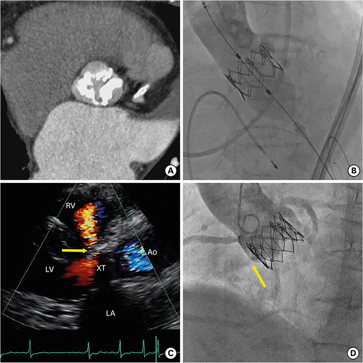
Figure 1 (A) Multislice computed tomography showing a severely calcified aortic valve with fusion of the right and non-coronary cusps. (B) A 26-mm SAPIEN XT (Edwards Lifesciences, Irvine, CA, USA) deployed via a transfemoral approach. (C) Transthoracic echocardiography showing atypical annulus rupture and a continuous shunt from the Ao to RV (arrow). (D) Aortography showing blood flow from the aortic annulus to RV (arrow). Ao = aorta; LA = left atrial; LV = left ventricle; RV = right ventricle.
Reference
-
1. Hayashida K, Bouvier E, Lefèvre T, et al. Potential mechanism of annulus rupture during transcatheter aortic valve implantation. Catheter Cardiovasc Interv. 2013; 82:E742–E746.
Article2. Hagiwara K, Saito N, Yamazaki K, Kimura T. Aorto-right ventricular fistula following transcatheter aortic valve implantation using a 29 mm SAPIEN XT valve. BMJ Case Rep. 2017; 2017:bcr-2017-219247.
Article
- Full Text Links
-
- Actions
-
Cited
- CITED
-
- Close
- Share
-
- Similar articles
-
- Expanding transcatheter aortic valve replacement into uncharted indications
- Transcatheter Mitral Valve Implantation in Open Heart Surgery: An Off-Label Technique
- Echocardiography in Transcatheter Aortic Valve Implantation and Mitral Valve Clip
- Similar Morphology, but Different Function: Acute Improvement of Myocardial Longitudinal Strain after Percutaneous Transcatheter Aortic Valve Implantation Therapy in a Severe Aortic Stenosis Patient
- Recent updates in transcatheter aortic valve implantation
