Impact of a Geometric Correction for Proximal Flow Constraint on the Assessment of Mitral Regurgitation Severity Using the Proximal Flow Convergence Method
- Affiliations
-
- 1Division of Cardiology, Department of Internal Medicine, Gyeongsang National University School of Medicine, Gyeongsang National University Changwon Hospital, Changwon, Korea.
- 2Cardiac Imaging Center, Asan Medical Center Heart Institute, University of Ulsan College of Medicine, Seoul, Korea. jksong@amc.seoul.kr
- 3Division of Cardiology, Department of Internal Medicine, Chungnam National University Hospital, Daejeon, Korea.
- KMID: 2407679
- DOI: http://doi.org/10.4250/jcu.2018.26.1.33
Abstract
- BACKGROUND
Overestimation of the severity of mitral regurgitation (MR) by the proximal isovelocity surface area (PISA) method has been reported. We sought to test whether angle correction (AC) of the constrained flow field is helpful to eliminate overestimation in patients with eccentric MR.
METHODS
In a total of 33 patients with MR due to prolapse or flail mitral valve, both echocardiography and cardiac magnetic resonance image (CMR) were performed to calculate regurgitant volume (RV). In addition to RV by conventional PISA (RV(PISA)), convergence angle (α) was measured from 2-dimensional Doppler color flow maps and RV was corrected by multiplying by α/180 (RV(AC)). RV measured by CMR (RV(CMR)) was used as a gold standard, which was calculated by the difference between total stroke volume measured by planimetry of the short axis slices and aortic stroke volume by phase-contrast image.
RESULTS
The correlation between RV(CMR) and RV by echocardiography was modest [RV(CMR) vs. RV(PISA) (r = 0.712, p < 0.001) and RV(CMR) vs. RV(AC) (r = 0.766, p < 0.001)]. However, RV(PISA) showed significant overestimation (RV(PISA) - RV(CMR) = 50.6 ± 40.6 mL vs. RV(AC) - RV(CMR) = 7.7 ± 23.4 mL, p < 0.001). The overall accuracy of RV(PISA) for diagnosis of severe MR, defined as RV ≥ 60 mL, was 57.6% (19/33), whereas it increased to 84.8% (28/33) by using RV(AC) (p = 0.028).
CONCLUSION
Conventional PISA method tends to provide falsely large RV in patients with eccentric MR and a simple geometric AC of the proximal constraint flow largely eliminates overestimation.
Keyword
MeSH Terms
Figure
-
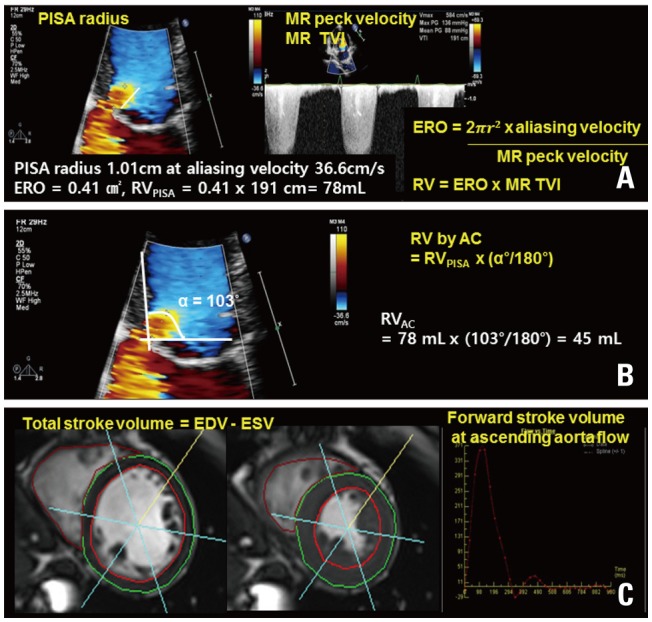
Fig. 1 A representative case showing the measurement of the RV using the conventional PISA method (A), the angle-corrected PISA (B), and cardiac magnetic resonance image (C). PISA: proximal isovelocity surface area, RV: regurgitant volume, AC: angle correction, MR: mitral regurgitation, EDV: end-diastolic volume, ESV: end-systolic volume, ERO: effective regurgitant orifice, TVI: time-velocity integral.
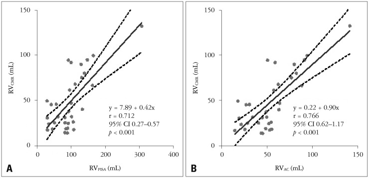
Fig. 2 Quantitative comparison of RV as determined by CMR (RVCMR) and echocardiography before (RVPISA) (A) and after the AC of the PISA method (RVAC) (B). CMR: cardiac magnetic resonance image, RV: regurgitant volume, PISA: proximal isovelocity surface area, AC: angle correction, CI: confidence interval.
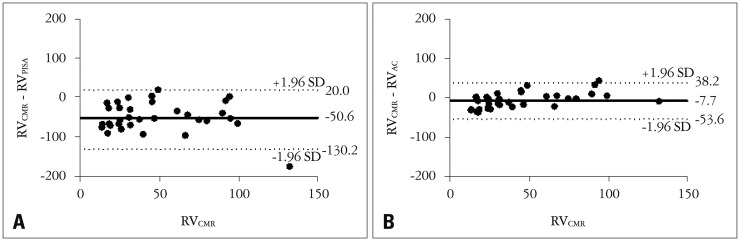
Fig. 3 Bland-Altman plot showing the difference of RV as determined by CMR (RVCMR) and echocardiography before (RVPISA) (A) and after the AC of the PISA method (RVAC) (B). CMR: cardiac magnetic resonance image, RV: regurgitant volume, PISA: proximal isovelocity surface area, AC: angle correction.
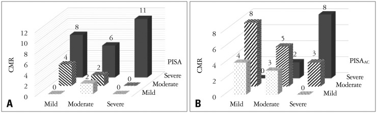
Fig. 4 Comparison of mitral regurgitation severity: CMR vs. conventional PISA (A) and CMR vs. angle-corrected PISA (B). CMR: cardiac magnetic resonance image, PISA: proximal isovelocity surface area, AC: angle correction.
Reference
-
1. Thavendiranathan P, Phelan D, Collier P, Thomas JD, Flamm SD, Marwick TH. Quantitative assessment of mitral regurgitation: how best to do it. JACC Cardiovasc Imaging. 2012; 5:1161–1175. PMID: 23153917.2. Lancellotti P, Moura L, Pierard LA, Agricola E, Popescu BA, Tribouilloy C, Hagendorff A, Monin JL, Badano L, Zamorano JL. European Association of Echocardiography. European Association of Echocardiography recommendations for the assessment of valvular regurgitation. Part 2: mitral and tricuspid regurgitation (native valve disease). Eur J Echocardiogr. 2010; 11:307–332. PMID: 20435783.3. Vahanian A, Alfieri O, Andreotti F, Antunes MJ, Barón-Esquivias G, Baumgartner H, Borger MA, Carrel TP, De Bonis M, Evangelista A, Falk V, Lung B, Lancellotti P, Pierard L, Price S, Schäfers HJ, Schuler G, Stepinska J, Swedberg K, Takkenberg J, Von Oppell UO, Windecker S, Zamorano JL, Zembala M. ESC Committee for Practice Guidelines (CPG). Joint Task Force on the Management of Valvular Heart Disease of the European Society of Cardiology (ESC). European Association for Cardio-Thoracic Surgery (EACTS). Guidelines on the management of valvular heart disease (version 2012): the joint task force on the management of valvular heart disease of the European Society of Cardiology (ESC) and the European Association for Cardio-Thoracic Surgery (EACTS). Eur J Cardiothorac Surg. 2012; 42:S1–S44. PMID: 22922698.4. Nishimura RA, Otto CM, Bonow RO, Carabello BA, Erwin JP 3rd, Guyton RA, O'Gara PT, Ruiz CE, Skubas NJ, Sorajja P, Sundt TM 3rd, Thomas JD. ACC/AHA Task Force Members. 2014 AHA/ACC guideline for the management of patients with valvular heart disease: executive summary: a report of the American College of Cardiology/American Heart Association Task Force on Practice Guidelines. Circulation. 2014; 129:2440–2492. PMID: 24589852.5. Moraldo M, Cecaro F, Shun-Shin M, Pabari PA, Davies JE, Xu XY, Hughes AD, Manisty C, Francis DP. Evidence-based recommendations for PISA measurements in mitral regurgitation: systematic review, clinical and in-vitro study. Int J Cardiol. 2013; 168:1220–1228. PMID: 23245796.6. Bellenger NG, Burgess MI, Ray SG, Lahiri A, Coats AJ, Cleland JG, Pennell DJ. Comparison of left ventricular ejection fraction and volumes in heart failure by echocardiography, radionuclide ventriculography and cardiovascular magnetic resonance; are they interchangeable? Eur Heart J. 2000; 21:1387–1396. PMID: 10952828.7. Biner S, Rafique A, Rafii F, Tolstrup K, Noorani O, Shiota T, Gurudevan S, Siegel RJ. Reproducibility of proximal isovelocity surface area, vena contracta, and regurgitant jet area for assessment of mitral regurgitation severity. JACC Cardiovasc Imaging. 2010; 3:235–243. PMID: 20223419.8. Uretsky S, Gillam L, Lang R, Chaudhry FA, Argulian E, Supariwala A, Gurram S, Jain K, Subero M, Jang JJ, Cohen R, Wolff SD. Discordance between echocardiography and MRI in the assessment of mitral regurgitation severity: a prospective multicenter trial. J Am Coll Cardiol. 2015; 65:1078–1088. PMID: 25790878.9. Pu M, Vandervoort PM, Griffin BP, Leung DY, Stewart WJ, Cosgrove DM 3rd, Thomas JD. Quantification of mitral regurgitation by the proximal convergence method using transesophageal echocardiography. Clinical validation of a geometric correction for proximal flow constraint. Circulation. 1995; 92:2169–2177. PMID: 7554198.10. Zoghbi WA, Enriquez-Sarano M, Foster E, Grayburn PA, Kraft CD, Levine RA, Nihoyannopoulos P, Otto CM, Quinones MA, Rakowski H, Stewart WJ, Waggoner A, Weissman NJ. American Society of Echocardiography. Recommendations for evaluation of the severity of native valvular regurgitation with two-dimensional and Doppler echocardiography. J Am Soc Echocardiogr. 2003; 16:777–802. PMID: 12835667.11. Chen C, Koschyk D, Brockhoff C, Heik S, Hamm C, Bleifeld W, Kupper W. Noninvasive estimation of regurgitant flow rate and volume in patients with mitral regurgitation by Doppler color mapping of accelerating flow field. J Am Coll Cardiol. 1993; 21:374–383. PMID: 8426001.12. Enriquez-Sarano M, Sinak LJ, Tajik AJ, Bailey KR, Seward JB. Changes in effective regurgitant orifice throughout systole in patients with mitral valve prolapse. A clinical study using the proximal isovelocity surface area method. Circulation. 1995; 92:2951–2958. PMID: 7586265.13. Gelfand EV, Hughes S, Hauser TH, Yeon SB, Goepfert L, Kissinger KV, Rofsky NM, Manning WJ. Severity of mitral and aortic regurgitation as assessed by cardiovascular magnetic resonance: optimizing correlation with Doppler echocardiography. J Cardiovasc Magn Reson. 2006; 8:503–507. PMID: 16755839.14. Kizilbash AM, Hundley WG, Willett DL, Franco F, Peshock RM, Grayburn PA. Comparison of quantitative Doppler with magnetic resonance imaging for assessment of the severity of mitral regurgitation. Am J Cardiol. 1998; 81:792–795. PMID: 9527098.15. Cawley PJ, Hamilton-Craig C, Owens DS, Krieger EV, Strugnell WE, Mitsumori L, D'Jang CL, Schwaegler RG, Nguyen KQ, Nguyen B, Maki JH, Otto CM. Prospective comparison of valve regurgitation quantitation by cardiac magnetic resonance imaging and transthoracic echocardiography. Circ Cardiovasc Imaging. 2013; 6:48–57. PMID: 23212272.16. Uretsky S, Supariwala A, Nidadovolu P, Khokhar SS, Comeau C, Shubayev O, Campanile F, Wolff SD. Quantification of left ventricular remodeling in response to isolated aortic or mitral regurgitation. J Cardiovasc Magn Reson. 2010; 12:32. PMID: 20497540.17. Acker MA, Parides MK, Perrault LP, Moskowitz AJ, Gelijns AC, Voisine P, Smith PK, Hung JW, Blackstone EH, Puskas JD, Argenziano M, Gammie JS, Mack M, Ascheim DD, Bagiella E, Moquete EG, Ferguson TB, Horvath KA, Geller NL, Miller MA, Woo YJ, D'Alessandro DA, Ailawadi G, Dagenais F, Gardner TJ, O'Gara PT, Michler RE, Kron IL. CTSN. Mitral-valve repair versus replacement for severe ischemic mitral regurgitation. N Engl J Med. 2014; 370:23–32. PMID: 24245543.18. Myerson SG, d'Arcy J, Christiansen JP, Dobson LE, Mohiaddin R, Francis JM, Prendergast B, Greenwood JP, Karamitsos TD, Neubauer S. Determination of clinical outcome in mitral regurgitation with cardiovascular magnetic resonance quantification. Circulation. 2016; 133:2287–2296. PMID: 27189033.19. Son JW, Chang HJ, Lee JK, Chung HJ, Song RY, Kim YJ, Datta S, Heo R, Shin SH, Cho IJ, Shim CY, Hong GR, Chung N. Automated quantification of mitral regurgitation by three dimensional real time full volume color Doppler transthoracic echocardiography: a validation with cardiac magnetic resonance imaging and comparison with two dimensional quantitative methods. J Cardiovasc Ultrasound. 2013; 21:81–89. PMID: 23837118.20. Thavendiranathan P, Liu S, Datta S, Walls M, Nitinunu A, Van Houten T, Tomson NA, Vidmar L, Georgescu B, Wang Y, Srinivasan S, De Michelis N, Raman SV, Ryan T, Vannan MA. Automated quantification of mitral inflow and aortic outflow stroke volumes by three-dimensional real-time volume color-flow Doppler transthoracic echocardiography: comparison with pulsed-wave Doppler and cardiac magnetic resonance imaging. J Am Soc Echocardiogr. 2012; 25:56–65. PMID: 22105057.21. Choi J, Heo R, Hong GR, Chang HJ, Sung JM, Shin SH, Cho IJ, Shim CY, Chung N. Differential effect of 3-dimensional color Doppler echocardiography for the quantification of mitral regurgitation according to the severity and characteristics. Circ Cardiovasc Imaging. 2014; 7:535–544. PMID: 24700692.22. Heo R, Son JW, Ó Hartaigh B, Chang HJ, Kim YJ, Datta S, Cho IJ, Shim CY, Hong GR, Ha JW, Chung N. Clinical implications of three-dimensional real-time color Doppler transthoracic echocardiography in quantifying mitral regurgitation: a comparison with conventional two-dimensional methods. J Am Soc Echocardiogr. 2017; 30:393–403.e7. PMID: 28238587.23. Thavendiranathan P, Liu S, Datta S, Rajagopalan S, Ryan T, Igo SR, Jackson MS, Little SH, De Michelis N, Vannan MA. Quantification of chronic functional mitral regurgitation by automated 3-dimensional peak and integrated proximal isovelocity surface area and stroke volume techniques using real-time 3-dimensional volume color Doppler echocardiography: in vitro and clinical validation. Circ Cardiovasc Imaging. 2013; 6:125–133. PMID: 23223636.24. Buck T, Plicht B. Real-time three-dimensional echocardiographic assessment of severity of mitral regurgitation using proximal isovelocity surface area and vena contracta area method. Lessons we learned and clinical implications. Curr Cardiovasc Imaging Rep. 2015; 8:38. PMID: 26322152.25. Hamada S, Altiok E, Frick M, Almalla M, Becker M, Marx N, Hoffmann R. Comparison of accuracy of mitral valve regurgitation volume determined by three-dimensional transesophageal echocardiography versus cardiac magnetic resonance imaging. Am J Cardiol. 2012; 110:1015–1020. PMID: 22727180.26. Thomas L, Foster E, Hoffman JI, Schiller NB. The mitral regurgitation index: an echocardiographic guide to severity. J Am Coll Cardiol. 1999; 33:2016–2022. PMID: 10362208.
- Full Text Links
-
- Actions
-
Cited
- CITED
-
- Close
- Share
-
- Similar articles
-
- Assessment of Mitral Valve Area in Patients with Mitral Stenosis Using Color Doppler Proximal Isovelocity Surface Area Method
- Quantitative Evaluation of Regurgitant Volume in the Patients with Mitral Regurgitation Using Color Doppler Proximal Isovelocity Surface Area Method
- A Comparative Study of the echocardiographic Methods Assessing Severity of Stenosis in Pateints with mitral Stenosis
- Color Doppler Assessment of Mitral Regurgitation
- Echocardiographic Assessment of Mitral Valve Regurgitation
