Optimization of scan delay for multi-phase computed tomography by using bolus tracking in normal canine kidney
- Affiliations
-
- 1College of Veterinary Medicine, Chonnam National University, Gwangju 61186, Korea. imsono@chonnam.ac.kr
- 2College of Veterinary Medicine, Chungbuk National University, Cheongju 28644, Korea.
- KMID: 2407628
- DOI: http://doi.org/10.4142/jvs.2018.19.2.290
Abstract
- This study was performed to optimize scan delays for canine kidney by using a bolus-tracking technique. In six beagle dogs, computed tomography (CT) of the kidney was performed three times in each dog with different scan delays after a bolus-tracking trigger of 100 Hounsfield units (HU) of aortic enhancement. Delays were 5, 20, 35, and 50 sec for the first scan, 10, 25, 40, and 55 sec for the second scan, and 15, 30, 45, and 60 sec for the third scan. The renal artery-to-vein contrast difference peaked at 5 sec, and the renal cortex-to-medulla contrast difference peaked at 10 sec. The renal cortex-to-medulla contrast difference approached zero at a scan delay of 30 sec after the bolus trigger. For the injection protocol used in this study, the optimal scan delay times for renal arterial, corticomedullary, and nephrographic phases were 5, 10, and 30 sec after triggering at 100 HU of aortic enhancement using the bolus-tracking technique. The bolus-tracking technique is useful in multi-phase renal CT study as it compensates for different transit times to the kidney among different animals, requires a small dose of contrast media, and does not require additional patient radiation exposure.
Keyword
Figure
-
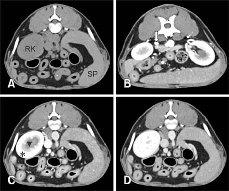
Fig. 1 Transverse computed tomography images with no enhancement (A), 5 sec scan delay (B), 10 sec scan delay (C), and 30 sec scan delay (D). (B) Renal arterial phase shows maximal renal artery contrast enhancement (arrow). (C) Corticomedullary phase shows the cortex (asterisk) enhances to a greater degree than the medulla (arrowhead) (D) Nephrographic phase shows a uniform renal enhancement. RK, right kidney; SP, spleen.
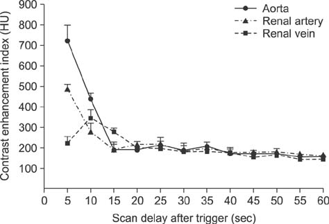
Fig. 2 Graph showing the scan delay after the bolus trigger vs. mean Hounsfield units (HU) for the abdominal aorta, renal artery, and renal vein.

Fig. 3 Graph showing the scan delay after the trigger vs. mean Hounsfield units (HU) for the renal cortex and medulla.
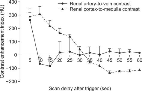
Fig. 4 Graph showing the scan delay after the bolus trigger of mean renal artery-to-vein contrast and renal cortex-to-medulla contrast. HU, Hounsfield units.
Reference
-
1. Bae KT, Heiken JP, Brink JA. Aortic and hepatic contrast medium enhancement at CT. Part II. Effect of reduced cardiac output in a porcine model. Radiology. 1998; 207:657–662.
Article2. Barthez PY, Begon D, Delisle F. Effect of contrast medium dose and image acquisition timing on ureteral opacification in the normal dog as assessed by computed tomography. Vet Radiol Ultrasound. 1998; 39:524–527.
Article3. Birnbaum BA, Jacobs JE, Langlotz CP, Ramchandani P. Assessment of a bolus-tracking technique in helical renal CT to optimize nephrographic phase imaging. Radiology. 1999; 211:87–94.
Article4. Birnbaum BA, Jacobs JE, Ramchandani P. Multiphasic renal CT: comparison of renal mass enhancement during the corticomedullary and nephrographic phases. Radiology. 1996; 200:753–758.
Article5. Bouma JL, Aronson LR, Keith DG, Saunders HM. Use of computed tomography renal angiography for screening feline renal transplant donors. Vet Radiol Ultrasound. 2003; 44:636–641.
Article6. Cáceres AV, Zwingenberger AL, Aronson LR, Mai W. Characterization of normal feline renal vascular anatomy with dual-phase CT angiography. Vet Radiol Ultrasound. 2008; 49:350–356.
Article7. Cohan RH, Sherman LS, Korobkin M, Bass JC, Francis IR. Renal masses: assessment of corticomedullary-phase and nephrographic-phase CT scans. Radiology. 1995; 196:445–451.
Article8. Dyer RB, Munitz HA, Bechtold R, Choplin RH. The abnormal nephrogram. Radiographics. 1986; 6:1039–1063.
Article9. Fields EL, Robertson ID, Brown JC Jr. Optimization of contrast-enhanced multidetector abdominal computed tomography in sedated canine patients. Vet Radiol Ultrasound. 2012; 53:507–512.
Article10. Goshima S, Kanematsu M, Nishibori H, Kondo H, Tsuge Y, Yokoyama R, Miyoshi T, Onozuka M, Shiratori Y, Moriyama N, Bae KT. Multi-detector row CT of the kidney: optimizing scan delays for bolus tracking techniques of arterial, corticomedullary, and nephrographic phases. Eur J Radiol. 2007; 63:420–426.
Article11. Hattery RR, Williamson B Jr, Hartman GW, LeRoy AJ, Witten DM. Intravenous urographic technique. Radiology. 1988; 167:593–599.
Article12. Heiken JP, Brink JA, Vannier MW. Spiral (helical) CT. Radiology. 1993; 189:647–656.
Article13. Herts BR, Coll DM, Novick AC, Obuchowski N, Linnell G, Wirth SL, Baker ME. Enhancement characteristics of papillary renal neoplasms revealed on triphasic helical CT of the kidneys. AJR Am J Roentgenol. 2002; 178:367–372.
Article14. Howard M. Clinical Urography. Philadelphia: W.B. Saunders;1990. p. 456–469.15. Kopka L, Fischer U, Zoeller G, Schmidt C, Ringert RH, Grabbe E. Dual-phase helical CT of the kidney: value of the corticomedullary and nephrographic phase for evaluation of renal lesions and preoperative staging of renal cell carcinoma. AJR Am J Roentgenol. 1997; 169:1573–1578.
Article16. Lee S, Jung J, Chang J, Yoon J, Choi M. Evaluation of triphasic helical computed tomography of the kidneys in clinically normal dogs. Am J Vet Res. 2011; 72:345–349.
Article17. Mai W, Suran JN, Cáceres AV, Reetz JA. Comparison between bolus tracking and timing-bolus techniques for renal computed tomographic angiography in normal cats. Vet Radiol Ultrasound. 2013; 54:343–350.
Article18. Prokop M. General principles of MDCT. Eur J Radiol. 2003; 45:Suppl 1. S4–S10.
Article19. Rankin SC, Webb JA, Reznek RH. Spiral computed tomography in the diagnosis of renal masses. BJU Int. 2000; 86:Suppl 1. 48–57.
Article20. Saunders HS, Dyer RB, Shifrin RY, Scharling ES, Bechtold RE, Zagoria RJ. The CT nephrogram: implications for evaluation of urinary tract disease. Radiographics. 1995; 15:1069–1085.
Article21. Schwarz T. Veterinary Computed Tomography. Ames: John Wiley & Sons;2013. p. 331.22. Silverman PM, Brown B, Wray H, Fox SH, Cooper C, Roberts S, Zeman RK. Optimal contrast enhancement of the liver using helical (spiral) CT: value of SmartPrep. AJR Am J Roentgenol. 1995; 164:1169–1171.
Article23. Szolar DH, Kammerhuber F, Altziebler S, Tillich M, Breinl E, Fotter R, Schreyer HH. Multiphasic helical CT of the kidney: increased conspicuity for detection and characterization of small (< 3-cm) renal masses. Radiology. 1997; 202:211–217.
Article24. Urban BA, Ratner LE, Fishman EK. Three-dimensional volume-rendered CT angiography of the renal arteries and veins: normal anatomy, variants, and clinical applications. Radiographics. 2001; 21:373–386.
Article25. Wisner ER. Atlas of Small Animal CT and MRI. Ames: John Wiley & Sons;2015. p. 584.26. Zeman RK, Zeiberg A, Hayes WS, Silverman PM, Cooper C, Garra BS. Helical CT of renal masses: the value of delayed scans. AJR Am J Roentgenol. 1996; 167:771–776.
Article
- Full Text Links
-
- Actions
-
Cited
- CITED
-
- Close
- Share
-
- Similar articles
-
- Optimal scan delay depending on contrast material injection duration in abdominal multi-phase computed tomography of pancreas and liver in normal Beagle dogs
- Variation of Attenuation Value of Pancreas at Dual Phase MDCT: Comparison of the Bolus-tracking Technique vs. the Fixed Scan Delay Protocol
- Values of Bolus Tracking Methods for Optimal Hepatic Enhancement
- Three-dimensional CT angiography of the canine hepatic vasculature
- Advantades of the intravenous bolus CT scan in differentiation of hepatic masses
