Pioglitazone Attenuates Palmitate-Induced Inflammation and Endoplasmic Reticulum Stress in Pancreatic β-Cells
- Affiliations
-
- 1Institute of Medical Research, Kangbuk Samsung Hospital, Sungkyunkwan University School of Medicine, Seoul, Korea.
- 2Division of Endocrinology and Metabolism, Department of Internal Medicine, Kangbuk Samsung Hospital, Sungkyunkwan University School of Medicine, Seoul, Korea. drlwy@hanmail.net
- KMID: 2407127
- DOI: http://doi.org/10.3803/EnM.2018.33.1.105
Abstract
- BACKGROUND
The nuclear receptor peroxisome proliferator-activator gamma (PPARγ) is a useful therapeutic target for obesity and diabetes, but its role in protecting β-cell function and viability is unclear.
METHODS
To identify the potential functions of PPARγ in β-cells, we treated mouse insulinoma 6 (MIN6) cells with the PPARγ agonist pioglitazone in conditions of lipotoxicity, endoplasmic reticulum (ER) stress, and inflammation.
RESULTS
Palmitate-treated cells incubated with pioglitazone exhibited significant improvements in glucose-stimulated insulin secretion and the repression of apoptosis, as shown by decreased caspase-3 cleavage and poly (adenosine diphosphate [ADP]-ribose) polymerase activity. Pioglitazone also reversed the palmitate-induced expression of inflammatory cytokines (tumor necrosis factor α, interleukin 6 [IL-6], and IL-1β) and ER stress markers (phosphor-eukaryotic translation initiation factor 2α, glucose-regulated protein 78 [GRP78], cleaved-activating transcription factor 6 [ATF6], and C/EBP homologous protein [CHOP]), and pioglitazone significantly attenuated inflammation and ER stress in lipopolysaccharide- or tunicamycin-treated MIN6 cells. The protective effect of pioglitazone was also tested in pancreatic islets from high-fat-fed KK-Ay mice administered 0.02% (wt/wt) pioglitazone or vehicle for 6 weeks. Pioglitazone remarkably reduced the expression of ATF6α, GRP78, and monocyte chemoattractant protein-1, prevented α-cell infiltration into the pancreatic islets, and upregulated glucose transporter 2 (Glut2) expression in β-cells. Moreover, the preservation of β-cells by pioglitazone was accompanied by a significant reduction of blood glucose levels.
CONCLUSION
Altogether, these results support the proposal that PPARγ agonists not only suppress insulin resistance, but also prevent β-cell impairment via protection against ER stress and inflammation. The activation of PPARγ might be a new therapeutic approach for improving β-cell survival and insulin secretion in patients with diabetes mellitus
Keyword
MeSH Terms
-
Animals
Apoptosis
Blood Glucose
Caspase 3
Chemokine CCL2
Cytokines
Diabetes Mellitus
Endoplasmic Reticulum Stress*
Endoplasmic Reticulum*
Glucose Transport Proteins, Facilitative
Humans
Inflammation*
Insulin
Insulin Resistance
Insulin-Secreting Cells
Insulinoma
Interleukin-6
Islets of Langerhans
Mice
Necrosis
Obesity
Peptide Initiation Factors
Peroxisomes
Repression, Psychology
Transcription Factors
Blood Glucose
Caspase 3
Chemokine CCL2
Cytokines
Glucose Transport Proteins, Facilitative
Insulin
Interleukin-6
Peptide Initiation Factors
Transcription Factors
Figure
-
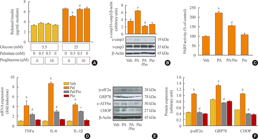
Fig. 1 Pioglitazone (Pio) improves palmitate (PA)-induced β-cell impairment via repression of the inflammatory response and endoplasmic reticulum (ER) stress. (A) Mouse insulinoma 6 (MIN6) cells were incubated with 0.5 mM PA in the presence or absence of 10 µM Pio for 24 hours, and the glucose-stimulated (1 or 4.5 g/L) insulin secretion of MIN6 cells was evaluated. Secreted insulin was measured by a mouse insulin enzyme-linked immunosorbent assay (ELISA) kit. The values are representative of 6 independent experiments. (B) Expression of cleaved caspase-3 (c-casp3), an apoptotic protein, was measured by Western blot analysis. (C) Poly (adenosine diphosphate [ADP]-ribose) polymerase (PARP) activity is represented as the percentage of relative absorbance compared to the vehicle group. (D) Transcription of tumor necrosis factor α (TNFA), interleukin 6 (IL6), and IL1B was measured by real time reverse-transcription polymerase chain reaction, and normalized with β-actin. (E) The ER stress proteins, including phosphor-eukaryotic translation initiation factor 2α (p-eIF2α), glucose-regulated protein 78 (GRP78), cleaved-activating transcription factor 6 (c-ATF6), and C/EBP homologous protein (CHOP), were measured by Western blot analysis and (F) the ratio of p-eIF2α, GRP78, and CHOP to β-actin was described. Each value represents the mean of three experiments. t-casp3, total caspase-3; Veh, vehicle; PA, palmitate. aP<0.01, bP<0.001 compared with the vehicle group; cP<0.05, dP<0.001 compared with the PA group.
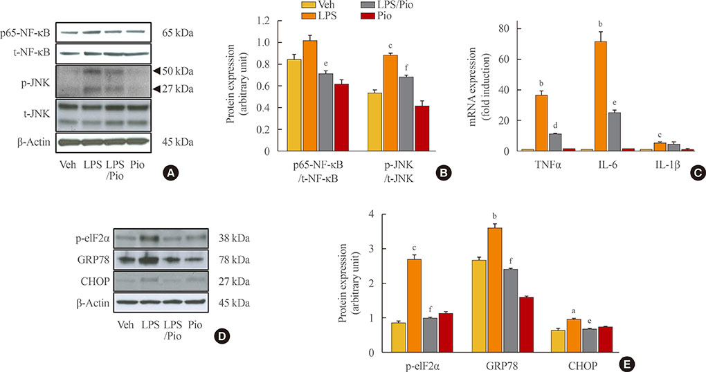
Fig. 2 Pioglitazone (Pio) represses lipopolysaccharide (LPS)-induced inflammatory response and endoplasmic reticulum (ER) stress. The mouse insulinoma 6 (MIN6) cells were incubated with 10 µg/mL LPS in the presence or absence of 10 µM Pio for 8 hours. (A) Phosphorylation of nuclear factor-kappa B (NF-κB) and jun N-terminal kinase (JNK), essential factors of the inflammatory response, was evaluated by Western blot. (B) The density of phosphorylated NF-κB and JNK was normalized to that of the total forms. (C) The inflammatory cytokines were measured by reverse-transcription polymerase chain reaction, and normalized with β-actin. (D) The ER stress proteins, including phosphor-eukaryotic translation initiation factor 2α (p-eIF2α), glucose-regulated protein 78 (GRP78), and C/EBP homologous protein (CHOP), were measured by Western blot analysis and (E) the ratio to β-actin was described. Each value represents the mean of three experiments. t-NF-κB, total nuclear factor kappa-light-chain-enhancer of activated B cells; p-JNK, phosphorylated c-Jun N-terminal kinase; t-JNK, total c-Jun N-terminal kinase; TNFα, tumor necrosis factor α; IL, interleukin. aP<0.05, bP<0.01, cP<0.001 compared with the vehicle (Veh) group; dP<0.05, eP<0.01, fP<0.001 compared with the LPS group.

Fig. 3 Pioglitazone (Pio) reduces endoplasmic reticulum (ER) stress, but not the inflammatory response in tunicamycin (TU)-challenged mouse insulinoma 6 (MIN6) cells. The MIN6 cells were incubated with 2 µg/mL TU in the presence or absence of 10 µM Pio for 24 hours. (A) The ER stress proteins, including phosphor-eukaryotic translation initiation factor 2α (p-eIF2α), glucose-regulated protein 78 (GRP78), cleaved-activating transcription factor 6 (c-ATF6), and C/EBP homologous protein (CHOP), were measured by Western blot analysis, and (B) the ratio to β-actin was described. (C) The transcription of tumor necrosis factor α (TNFA), interleukin 6 (IL6), and IL1B was measured by real-time reverse-transcription polymerase chain reaction, and normalized with β-actin. Each value represents the mean of three experiments. aP<0.05, bP<0.01, cP<0.001 compared with the vehicle (Veh) group; dP<0.01, eP<0.001 compared with the TU group.
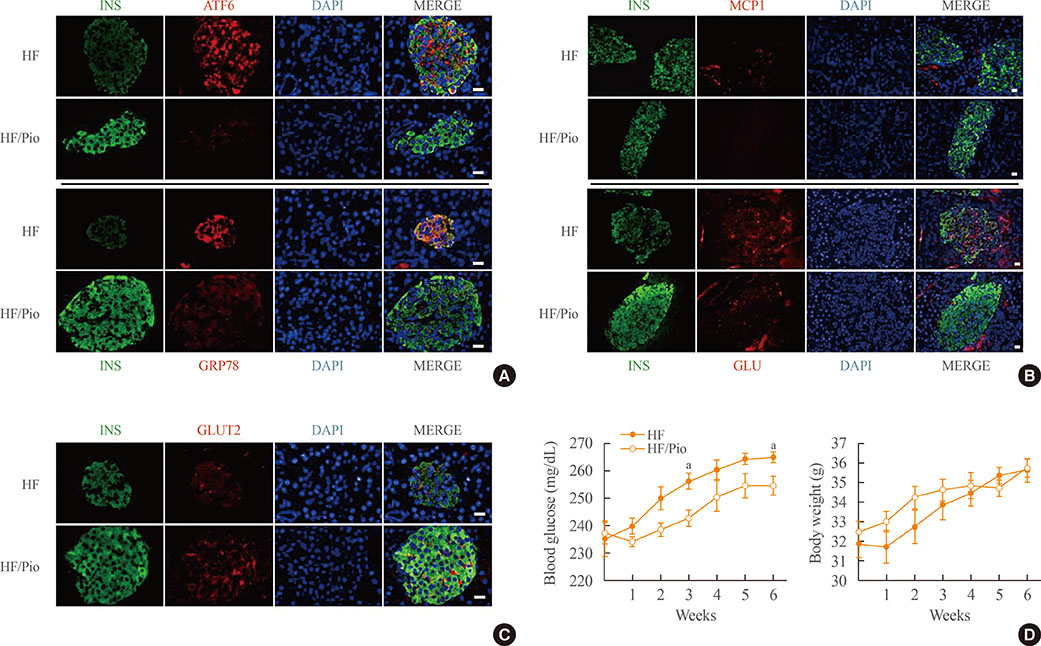
Fig. 4 Pioglitazone (Pio) protects pancreatic β-cells and regulates blood glucose levels in high-fat (HF)-diet-induced diabetic mice. The pancreatic islet from KK-Ay mice, which were fed an HF diet with or without Pio, were examined by double-immunofluorescence for (A) insulin-activating transcription factor 6 and insulin-glucose-regulated protein 78 (GRP78), (B) insulin-monocyt e chemoattractant protein-1 and insulin-glucagon (GLU), (C) insulin-glucose transporter 2 (GLUT2). (A-C) The nucleus was visualized by 4′,6-diamidino-2-phenylindole (DAPI). Bars=20 µm. (D) Blood glucose levels and body weight were measured every week. INS, insulin; ATF6, activating transcription factor 6; MCP1, monocyte chemoattractant protein-1. aP<0.05 compared with the HF group.
Cited by 2 articles
-
Inflammation in Metabolic Diseases and Insulin Resistance
Won-Young Lee
Cardiovasc Prev Pharmacother. 2021;3(2):31-37. doi: 10.36011/cpp.2021.3.e5.Targets for rescue from fatty acid-induced lipotoxicity in pancreatic beta cells
Seok-Woo Hong, Won-Young Lee
Cardiovasc Prev Pharmacother. 2022;4(2):57-62. doi: 10.36011/cpp.2022.4.e9.
Reference
-
1. Poitout V, Amyot J, Semache M, Zarrouki B, Hagman D, Fontes G. Glucolipotoxicity of the pancreatic beta cell. Biochim Biophys Acta. 2010; 1801:289–298.
Article2. Ishida H, Takizawa M, Ozawa S, Nakamichi Y, Yamaguchi S, Katsuta H, et al. Pioglitazone improves insulin secretory capacity and prevents the loss of beta-cell mass in obese diabetic db/db mice: possible protection of beta cells from oxidative stress. Metabolism. 2004; 53:488–494.3. Lin CY, Gurlo T, Haataja L, Hsueh WA, Butler PC. Activation of peroxisome proliferator-activated receptor-gamma by rosiglitazone protects human islet cells against human islet amyloid polypeptide toxicity by a phosphatidylinositol 3’-kinase-dependent pathway. J Clin Endocrinol Metab. 2005; 90:6678–6686.4. Kim HS, Hwang YC, Koo SH, Park KS, Lee MS, Kim KW, et al. PPAR-γ activation increases insulin secretion through the up-regulation of the free fatty acid receptor GPR40 in pancreatic β-cells. PLoS One. 2013; 8:e50128.
Article5. Montane J, Cadavez L, Novials A. Stress and the inflammatory process: a major cause of pancreatic cell death in type 2 diabetes. Diabetes Metab Syndr Obes. 2014; 7:25–34.6. Larsen CM, Faulenbach M, Vaag A, Volund A, Ehses JA, Seifert B, et al. Interleukin-1-receptor antagonist in type 2 diabetes mellitus. N Engl J Med. 2007; 356:1517–1526.
Article7. Cai K, Qi D, Wang O, Chen J, Liu X, Deng B, et al. TNF-α acutely upregulates amylin expression in murine pancreatic beta cells. Diabetologia. 2011; 54:617–626.
Article8. Montane J, Klimek-Abercrombie A, Potter KJ, Westwell-Roper C, Bruce Verchere C. Metabolic stress, IAPP and islet amyloid. Diabetes Obes Metab. 2012; 14:Suppl 3. 68–77.
Article9. Masters SL, Dunne A, Subramanian SL, Hull RL, Tannahill GM, Sharp FA, et al. Activation of the NLRP3 inflammasome by islet amyloid polypeptide provides a mechanism for enhanced IL-1β in type 2 diabetes. Nat Immunol. 2010; 11:897–904.10. van Raalte DH, Diamant M. Glucolipotoxicity and beta cells in type 2 diabetes mellitus: target for durable therapy? Diabetes Res Clin Pract. 2011; 93:Suppl 1. S37–S46.
Article11. Igoillo-Esteve M, Marselli L, Cunha DA, Ladriere L, Ortis F, Grieco FA, et al. Palmitate induces a pro-inflammatory response in human pancreatic islets that mimics CCL2 expression by beta cells in type 2 diabetes. Diabetologia. 2010; 53:1395–1405.
Article12. Nakatani Y, Kaneto H, Kawamori D, Yoshiuchi K, Hatazaki M, Matsuoka TA, et al. Involvement of endoplasmic reticulum stress in insulin resistance and diabetes. J Biol Chem. 2005; 280:847–851.
Article13. Marchetti P, Bugliani M, Lupi R, Marselli L, Masini M, Boggi U, et al. The endoplasmic reticulum in pancreatic beta cells of type 2 diabetes patients. Diabetologia. 2007; 50:2486–2494.
Article14. Laybutt DR, Preston AM, Akerfeldt MC, Kench JG, Busch AK, Biankin AV, et al. Endoplasmic reticulum stress contributes to beta cell apoptosis in type 2 diabetes. Diabetologia. 2007; 50:752–763.
Article15. Li G, Mongillo M, Chin KT, Harding H, Ron D, Marks AR, et al. Role of ERO1-alpha-mediated stimulation of inositol 1,4,5-triphosphate receptor activity in endoplasmic reticulum stress-induced apoptosis. J Cell Biol. 2009; 186:783–792.16. Liu H, Cao MM, Wang Y, Li LC, Zhu LB, Xie GY, et al. Endoplasmic reticulum stress is involved in the connection between inflammation and autophagy in type 2 diabetes. Gen Comp Endocrinol. 2015; 210:124–129.
Article17. Donath MY, Boni-Schnetzler M, Ellingsgaard H, Ehses JA. Islet inflammation impairs the pancreatic beta-cell in type 2 diabetes. Physiology (Bethesda). 2009; 24:325–331.18. Finegood DT, McArthur MD, Kojwang D, Thomas MJ, Topp BG, Leonard T, et al. Beta-cell mass dynamics in Zucker diabetic fatty rats. Rosiglitazone prevents the rise in net cell death. Diabetes. 2001; 50:1021–1029.19. DREAM (Diabetes REduction Assessment with ramipril and rosiglitazone Medication) Trial Investigators. Gerstein HC, Yusuf S, Bosch J, Pogue J, Sheridan P, et al. Effect of rosiglitazone on the frequency of diabetes in patients with impaired glucose tolerance or impaired fasting glucose: a randomised controlled trial. Lancet. 2006; 368:1096–1105.20. Higa M, Zhou YT, Ravazzola M, Baetens D, Orci L, Unger RH. Troglitazone prevents mitochondrial alterations, beta cell destruction, and diabetes in obese prediabetic rats. Proc Natl Acad Sci U S A. 1999; 96:11513–11518.
Article21. Dubois M, Pattou F, Kerr-Conte J, Gmyr V, Vandewalle B, Desreumaux P, et al. Expression of peroxisome proliferator-activated receptor gamma (PPARgamma) in normal human pancreatic islet cells. Diabetologia. 2000; 43:1165–1169.22. Lazar MA. PPAR gamma, 10 years later. Biochimie. 2005; 87:9–13.23. Rosen ED, Kulkarni RN, Sarraf P, Ozcan U, Okada T, Hsu CH, et al. Targeted elimination of peroxisome proliferator-activated receptor gamma in beta cells leads to abnormalities in islet mass without compromising glucose homeostasis. Mol Cell Biol. 2003; 23:7222–7229.24. Unger RH, Zhou YT, Orci L. Regulation of fatty acid homeostasis in cells: novel role of leptin. Proc Natl Acad Sci U S A. 1999; 96:2327–2332.
Article25. Pahan K, Sheikh FG, Khan M, Namboodiri AM, Singh I. Sphingomyelinase and ceramide stimulate the expression of inducible nitric-oxide synthase in rat primary astrocytes. J Biol Chem. 1998; 273:2591–2600.
Article26. Maedler K, Sergeev P, Ris F, Oberholzer J, Joller-Jemelka HI, Spinas GA, et al. Glucose-induced beta cell production of IL-1beta contributes to glucotoxicity in human pancreatic islets. J Clin Invest. 2002; 110:851–860.27. Bernal-Mizrachi E, Wen W, Shornick M, Permutt MA. Activation of nuclear factor-kappaB by depolarization and Ca(2+) influx in MIN6 insulinoma cells. Diabetes. 2002; 51:Suppl 3. S484–S488.28. Lawrence T. The nuclear factor NF-kappaB pathway in inflammation. Cold Spring Harb Perspect Biol. 2009; 1:a001651.29. Kono T, Ahn G, Moss DR, Gann L, Zarain-Herzberg A, Nishiki Y, et al. PPAR-γ activation restores pancreatic islet SERCA2 levels and prevents β-cell dysfunction under conditions of hyperglycemic and cytokine stress. Mol Endocrinol. 2012; 26:257–271.
Article
- Full Text Links
-
- Actions
-
Cited
- CITED
-
- Close
- Share
-
- Similar articles
-
- Endoplasmic Reticulum Stress and Diabetes
- Metformin Ameliorates Lipotoxic β-Cell Dysfunction through a Concentration-Dependent Dual Mechanism of Action
- Endoplasmic Reticulum Stress and Dysregulated Autophagy in Human Pancreatic Beta Cells
- Carbon monoxide releasing molecule-2 protects mice against acute kidney injury through inhibition of ER stress
- AM1638, a GPR40-Full Agonist, Inhibited Palmitate- Induced ROS Production and Endoplasmic Reticulum Stress, Enhancing HUVEC Viability in an NRF2-Dependent Manner
