Smooth Muscle Tumor of Uncertain Malignant Potential Originating from the Superior Mesenteric Vein: A Case Report
- Affiliations
-
- 1Department of Radiology, Myongji Hospital, Seonam University College of Medicine, Goyang, Korea.
- 2Department of Radiology, Kangbuk Samsung Hospital, Sungkyunkwan University School of Medicine, Seoul, Korea. carukeion@hanmail.net
- 3Department of Pathology, Myongji Hospital, Seonam University College of Medicine, Goyang, Korea.
- KMID: 2405735
- DOI: http://doi.org/10.3348/jksr.2017.77.3.192
Abstract
- Uterine smooth muscle neoplasms that cannot be clearly diagnosed as benign or malignant based on generally applied histopathologic parameters are classified as "smooth muscle tumors of uncertain malignant potential (STUMP)". Herein, we report on the first case of a STUMP that originated from the superior mesenteric vein (SMV), with emphasis on the radiologic findings. Magnetic resonance imaging showed a well-defined mass encasing the SMV with progressive and homogeneous enhancement in the arterial and portal venous phases. The lesion showed mild hyperintensity on T2-weighted images when compared with skeletal muscle, which was quite different from typical leiomyomas. The lesion also showed hyperintensity on the diffusion-weighted images, and hypointensity on the apparent diffusion coefficient map. These results may reflect the high cellularity of the mass.
MeSH Terms
Figure
-
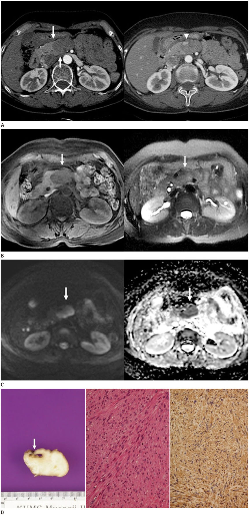
Fig. 1 Smooth muscle tumor of uncertain malignant potential originating from the SMV in a 59-year-old woman. A. On axial contrast-enhanced CT images, an approximately 4 × 3 × 3 cm sized, well-defined mass showed progressively homogeneous enhancement during arterial (left) and portal phases (right). The pancreas head appears compressed by the lesion with a sharp, beak-shaped interface (arrows). The mass is encasing the entire circumference of the SMV (arrowhead). B. On MR images, the mass shows slightly low signal intensity (arrow) on a T1-weighted image (left) and slightly high signal intensity (arrow) on a T2-weighted image (right) compared with that of skeletal muscle. Gadoxetic acid-enhanced MR images revealed progressive homogeneous enhancement (not shown). SMV = superior mesenteric vein C. A diffusion-weighted image obtained with a b value of 600 sec/mm2 (left) shows hyperintensity (arrow), and the apparent diffusion coefficient map (right) shows hypointensity of the mass (arrow). D. Grossly (left), the cut surface reveals a firm and circumscribed grayish white mass encircling the SMV (arrow). Microscopically, spindle cell proliferation with moderate cytologic atypia and no tumor necrosis (hematoxylin and eosin stain, × 200, middle) was shown. Immunohistochemical staining for SMA (× 200, right) shows diffuse positivity. SMA = smooth muscle actin, SMV = superior mesenteric vein
Reference
-
1. Fletcher CDM, Unni KK, Mertens F. World Health Organization classification of tumors: pathology and genetics of tumors of soft tissue and bone. Lyon: IARC Press;2002.2. Nishino M, Hayakawa K, Minami M, Yamamoto A, Ueda H, Takasu K. Primary retroperitoneal neoplasms: CT and MR imaging findings with anatomic and pathologic diagnostic clues. Radiographics. 2003; 23:45–57.3. Bell SW, Kempson RL, Hendrickson MR. Problematic uterine smooth muscle neoplasms. A clinicopathologic study of 213 cases. Am J Surg Pathol. 1994; 18:535–558.4. Won HS, Chun HG, Lee K. Retroperitoneal smooth muscle tumor of uncertain malignant potential after hysterectomy: a case report. J Med Case Rep. 2011; 5:214.5. Ueda H, Togashi K, Konishi I, Kataoka ML, Koyama T, Fujiwara T, et al. Unusual appearances of uterine leiomyomas: MR imaging findings and their histopathologic backgrounds. Radiographics. 1999; 19 Spec No:S131–S145.6. Yamashita Y, Torashima M, Takahashi M, Tanaka N, Katabuchi H, Miyazaki K, et al. Hyperintense uterine leiomyoma at T2-weighted MR imaging: differentiation with dynamic enhanced MR imaging and clinical implications. Radiology. 1993; 189:721–725.7. Tanaka YO, Nishida M, Tsunoda H, Okamoto Y, Yoshikawa H. Smooth muscle tumors of uncertain malignant potential and leiomyosarcomas of the uterus: MR findings. J Magn Reson Imaging. 2004; 20:998–1007.8. Koh DM, Collins DJ. Diffusion-weighted MRI in the body: applications and challenges in oncology. AJR Am J Roentgenol. 2007; 188:1622–1635.9. Ip PP, Cheung AN, Clement PB. Uterine smooth muscle tumors of uncertain malignant potential (STUMP): a clinicopathologic analysis of 16 cases. Am J Surg Pathol. 2009; 33:992–1005.
- Full Text Links
-
- Actions
-
Cited
- CITED
-
- Close
- Share
-
- Similar articles
-
- A Case of Cutaneous Smooth Muscle Tumor of Uncertain Malignant Potential
- A Clinical - Pathological Study of Uterine Smooth Muscle Tumor of Uncertain Malignant Potential
- A case of retroperitoneal huge smooth muscle tumor misleading to ovarian cancer
- Retroperitoneal recurrence of uterine smooth muscle tumor of uncertain malignant potential as leiomyosarcoma
- Acute Appendicitis with Superior Mesenteric Vein Thrombosis and Portal Vein Thrombosis
