Primary Presacral Neuroendocrine Tumor: A Case Report and Review of MRI Findings
- Affiliations
-
- 1Department of Radiology, Pusan National University Hospital, Pusan National University School of Medicine, Busan, Korea. leenk77@hanmail.net
- 2Department of Pathology, Pusan National University Hospital, Pusan National University School of Medicine, Busan, Korea.
- 3Department of Obstetrics and Gynecology, Pusan National University Hospital, Pusan National University School of Medicine, Busan, Korea.
- KMID: 2405734
- DOI: http://doi.org/10.3348/jksr.2017.77.3.187
Abstract
- Primary presacral neuroendocrine tumor (NET) is extremely rare. Furthermore, its preoperative diagnosis is very difficult, and its imaging characteristics are not well described. We report the case of a 70-year-old female with presacral NET, and describe its imaging features on diffusion-weighted magnetic resonance imaging.
MeSH Terms
Figure
-
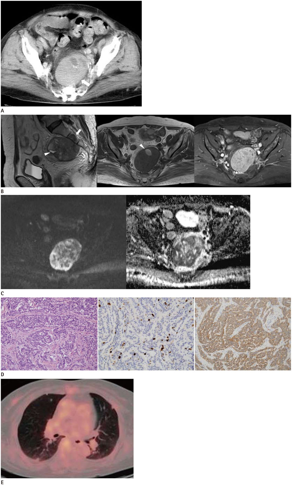
Fig. 1 A 70-year-old woman presented with primary presacral neuroendocrine tumor. A. Contrast-enhanced CT reveals an 8 cm-sized presacral mass (arrow) which abutted the sacrum without bony erosion or destruction. The mass is well-defined, solid with heterogeneous enhancement and it shows internal cystic degeneration and calcification. B. Sagittal T2-weighted image (left) shows a presacral mass with pressure erosion and bone marrow change (arrow) in the sacrum. The mass shows heterogeneous intermediate signal intensity on T2-weighted image (left), hypointensity on T1-weighted image (middle), and avid enhancement on contrast-enhanced T1-weighted image (right). Focal hyperintense foci (arrowheads) within the mass are noted on T1- and T2-weighted images. C. The tumor exhibits hyperintensity on diffusion-weighted image (left) at a b value of 1000 s/mm2 with a low apparent diffusion coefficient (ADC) value (mean, 0.632 × 10⁻³ mm2/sec) on the ADC map (right). D. Photomicrograph of the tumor cells (left, hematoxylin and eosin stain, × 200) shows an infiltrative border in the background of fibrotic stroma. Tumor shows monomorphic, plasmacytoid neuroendocrine cells, with insular, solid and trabecular growth patterns. Immunohistochemical examinations show approximately 15% increase in the Ki-67 labeling index (middle, × 400) and positivity for synaptophysin (right, × 200). E. Positron emission tomography-CT shows multiple pulmonary nodules, which are suspicious for metastasis.
Reference
-
1. Dujardin F, Beaussart P, de Muret A, Rosset P, Waynberger E, Mulleman D, et al. Primary neuroendocrine tumor of the sacrum: case report and review of the literature. Skeletal Radiol. 2009; 38:819–823.2. Klimstra DS, Modlin IR, Coppola D, Lloyd RV, Suster S. The pathologic classification of neuroendocrine tumors: a review of nomenclature, grading, and staging systems. Pancreas. 2010; 39:707–712.3. Hain KS, Pickhardt PJ, Lubner MG, Menias CO, Bhalla S. Presacral masses: multimodality imaging of a multidisciplinary space. Radiographics. 2013; 33:1145–1167.4. Lee JE, Shin KS, Cho JS, Kang DY, Kim JY. Presacral primary well differentiated neuroendocrine carcinoma: case report. J Korean Soc Radiol. 2011; 64:475–479.5. Kim T, Grobmyer SR, Liu C, Hochwald SN. Primary presacral neuroendocrine tumor associated with imperforate anus. World J Surg Oncol. 2007; 5:115.6. Misawa S, Horie H, Yamaguchi T, Kobayashi S, Kumano H, Lefor AT, et al. A unique retrorectal tumor with neuroendocrine differentiation: case report and review of the literature. Int J Surg Pathol. 2013; 21:271–277.7. Wong JFS, Teo CCM. An unusual case of presacral carcinoid tumor and the approach of management. Am J Cancer Case Rep. 2013; 1:21–26.8. Sahani DV, Bonaffini PA, Fernández-Del Castillo C, Blake MA. Gastroenteropancreatic neuroendocrine tumors: role of imaging in diagnosis and management. Radiology. 2013; 266:38–61.9. Kim JH, Eun HW, Kim YJ, Han JK, Choi BI. Staging accuracy of MR for pancreatic neuroendocrine tumor and imaging findings according to the tumor grade. Abdom Imaging. 2013; 38:1106–1114.
- Full Text Links
-
- Actions
-
Cited
- CITED
-
- Close
- Share
-
- Similar articles
-
- Presacral Primary Well Differentiated Neuroendocrine Carcinoma: Case Report
- MRI Findings of an Ampulla of Vater Neuroendocrine Tumor with Liver and Lymph Node Metastasis: a Case Report
- Primary Small Cell Neuroendocrine Carcinoma of the Breast: A Case Report With Literature Review
- Primary Neuroendocrine Carcinoma of the Breast: A Case Report and Literature Review
- Mammographic, Sonographic, and MRI Features of Primary Neuroendocrine Carcinoma of the Breast: A Case Report
