Esthetic implant restoration in the maxillary anterior missing area with palatal defect of the alveolar bone: a case report
- Affiliations
-
- 1Department of Prosthodontics, School of Dentistry and Institute of Oral Bio-Science, Chonbuk National University, Jeonju, Republic of Korea. jmseo@jbnu.ac.kr
- 2Department of Dentistry, School of Medicine, Eulji University, Daejeon, Republic of Korea.
- KMID: 2405542
- DOI: http://doi.org/10.14368/jdras.2017.33.4.291
Abstract
- It is challenging to produce esthetic implant restoration in the narrow anterior maxilla region where insufficient volume of alveolar bone could limit the angle and position of implant fixture, if preceding bone augmentation is not considered. Ideal angle and position of implant fixture placement should be established to reproduce harmonious emergence profile with marginal gingiva of implant prosthesis, bone augmentation considered to be preceded before implant placement occasionally. In this case, preceding bone augmentation has been operated before esthetic implant prosthesis in narrow anterior maxilla region. Preceded excessive bone augmentation in buccal area allowed proper angulation of implantation, which compensates unfavorable implant position. Provisional restorations were corrected during sufficient period to make harmonious level of marginal gingiva and interdental papilla. The definite restoration was fabricated using zirconia core based glass ceramic. Functionally and esthetically satisfactory results were obtained.
Keyword
Figure
-
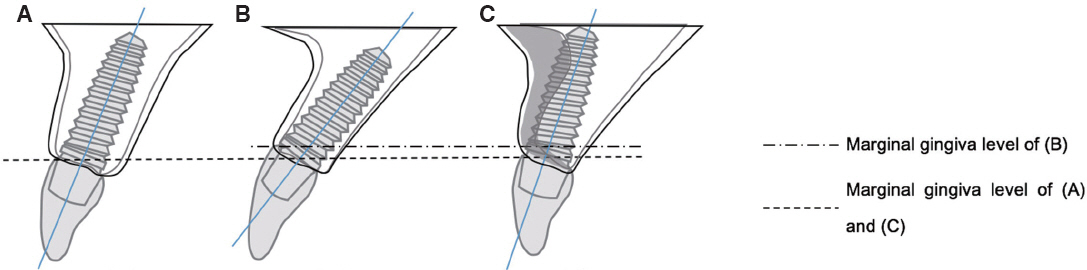
Fig. 1 Position and angulation of implant fixture under volume of alveolar bone. (A) Ideal formation of implantation, (B) Angulated fixture may emerge from too buccal position of alveolar bone, (C) Implant fixture can be located in ideal position of alveolar bone after preceded bone augmentation

Fig. 2 (A) Intraoral photograph at first visit, (B) Periapical standard images at first visit
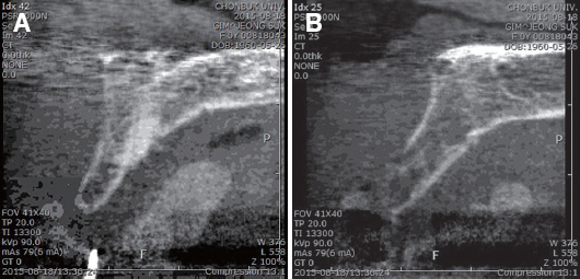
Fig. 3 Coronal CT images of edentulous area. (A) #21 area, (B) #22 area
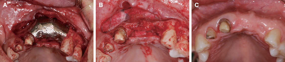
Fig. 4 Bone augmentation. (A) Narrow edentulous ridge of operation site: before operation, (B) Flap elevation: thin width with palatal defect of alveolar bone, (C) After applying of bone graft material and Ti-mesh
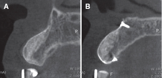
Fig. 5 CT images 3 months after bone augmentation. (A) #21 area, (B) #22 area

Fig. 6 Implantation on #21, 22 area. (A) Occlusal view, (B) Labial view
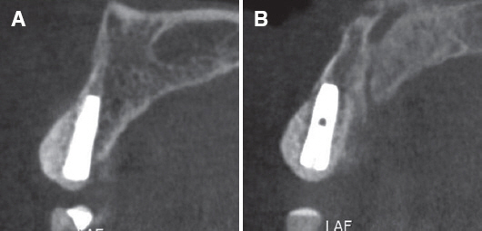
Fig. 7 CT images 4 months after implantation; fixture placed tilted to the palatal side. (A) #21 area, (B) #22 area
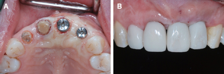
Fig. 8 After 2nd surgery using punch technique. (A) Intraoral image of operation site after connecting healing abutment, (B) Front view of provisional restoration

Fig. 9 Intraoral images 3 months after 2nd surgery. (A) Frontal view, (B) Left lateral view
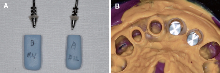
Fig. 10 Final impression using pick-up impression coping. (A) Duplicated emergence profile of provisional prosthesis on pick-up impression coping, (B) Impression using individualized impression coping and silicon impression material

Fig. 11 Intraoral images after connection of individualized zirconia abutment. (A) Frontal view, (B) Occlusal view
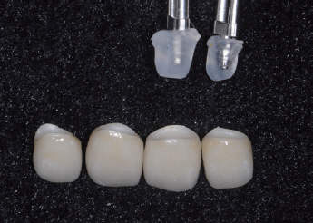
Fig. 12 Abutment replica using hot melt adhesive to minimize residual cement

Fig. 13 Intraoral images after final setting with resin cement. (A) Frontal view, (B) Left lateral view
Reference
-
References
1. Buser D, Martin W, Belser UC. Optimizing esthetics for implant restorations in the anterior maxilla: anatomic and surgical considerations. Int J Oral Maxillofac Implants. 2004; 19:43–61. PMID: 15635945.2. Seo CW, Han AR, Seo JM, Lee JJ. A technique for fabricating abutment replica with hot melt adhesive material to minimize residual cement in implant restoration: a case report. J Dent Rehabil Appl Sci. 2016; 32:240–5. DOI: 10.14368/jdras.2016.32.3.240.3. Araújo MG, Lindhe J. Dimensional ridge alterations following tooth extraction. An experimental study in the dog. J Clin Periodontol. 2015; 32:212–8. DOI: 10.1111/j.1600-051X.2005.00642.x. PMID: 15691354.4. Malchiodi L, Scarano A, Quaranta M, Piattelli A. Rigid fixation by means of titanium mesh in edentulous ridge expansion for horizontal ridge augmentation in the maxilla. Int J Oral Maxillofac Implants. 1998; 13:701–5. PMID: 9796156.5. von Arx T, Hardt N, Wallkamm B. The TIME technique: a new method for localized alveolar ridge augmentation prior to placement of dental implants. Int J Oral Maxillofac Implants. 1996; 11:387–94. PMID: 8752560.6. von Arx T, Kurt B. Implant placement and simultaneous ridge augmentation using autogenous bone and a micro titanium mesh: a prospective clinical study with 20 implants. Clin Oral Implants Res. 1999; 10:24–33. DOI: 10.1034/j.1600-0501.1999.100104.x.7. Phillips JH, Rahn BA. Fixation effects on membranous and endochondral onlay bone graft revascularization and bone deposition. Plast Reconstr Surg. 1990; 85:891–7. DOI: 10.1097/00006534-199006000-00009.8. Zins JE, Whitaker LA. Membranous versus endochondral bone: implications for craniofacial reconstruction. Plast Reconstr Surg. 1983; 72:778–85. DOI: 10.1097/00006534-198312000-00005.9. Rabie AB, Dan Z, Samman N. Ultrastructural identification of cells involved in the healing of intramembraneous and endochondral bones. Int J Oral Maxillofac Surg. 1996; 25:383–8. DOI: 10.1016/S0901-5027(06)80038-X.10. Roccuzzo M, Ramieri G, Spada MC, Bianchi SD, Berrone S. Vertical alveolar ridge augmentation by means of a titanium mesh and autogenous bone grafts. Clin Oral Implants Res. 2004; 15:73–81. DOI: 10.1111/j.1600-0501.2004.00998.x. PMID: 14731180.11. Lazzara RJ. Managing the soft tissue margin: the key to implant aesthetics. Pract Periodontics Aesthet Dent. 1993; 5:81–8. PMID: 8219171.12. Hürzeler MB, Quiñones CR, Strub JR. Advanced surgical and prosthetic management of the anterior single tooth osseointegrated implant: a case presentation. Pract Periodontics Aesthet Dent. 1994; 6:13–21. PMID: 8180367.13. Bichacho N, Landsberg CJ. A modified surgical/prosthetic approach for an optimal single implantsupported crown. Part II. The cervical contouring concept. Pract Periodontics Aesthet Dent. 1994; 6:35–41. PMID: 8054640.14. Grunder U. Stability of the mucosal topography around single-tooth implants and adjacent teeth:1-year results. Int J Periodontics Restorative Dent. 2000; 20:11–7. PMID: 11203544.15. Small PN, Tarnow DP. Gingival recession around implants: a 1-year longitudinal prospective study. Int J Oral Maxillofac Implants. 2000; 15:527–32. PMID: 10960986.16. Funato A, Salama MA, Ishikawa T, Garber DA, Salama H. Timing, positioning, and sequential staging in esthetic implant therapy: a four-dimensional perspective. Int J Periodontics Restorative Dent. 2007; 27:313–23. PMID: 17726987.17. Misch CE. Contemporary implant dentistry. 3rd ed. St. Louis: CV Mosby;2009. p. 739–68.18. Bengazi F, Wennström JL, Lekholm U. Recession of the soft tissue margin at oral implants. A 2-year longitudinal prospective study. Clin Oral Implants Res. 1996; 7:303–10. DOI: 10.1034/j.1600-0501.1996.070401.x. PMID: 9151595.
- Full Text Links
-
- Actions
-
Cited
- CITED
-
- Close
- Share
-
- Similar articles
-
- Cases of screw-retained implant prosthesis in the anterior maxilla through multidisciplinary approach, including orthodontic teeth alignment
- Ridge Augmentation Using Vascularized Interpositional Periosteal- Connective Tissue (VIP-CT) in Conjunction with Anterior Implant Placement in Maxilla: Report of Three Cases
- Esthetic restoration in continuous maxillary anterior area using immediate implant placement: A case report
- Single-tooth implant restoration with alveolar bone augmentation in the maxillary anterior tooth region: a case report
- Anterior esthetic restoration using DSD (digital smile design) for a patient with congenital missing tooth of maxillary central incisor
