Plug-Assisted Retrograde Transvenous Obliteration for the Treatment of Gastric Varices: The Role of Intra-Procedural Cone-Beam Computed Tomography
- Affiliations
-
- 1Department of Radiology and Research Institute of Radiology, University of Ulsan College of Medicine, Asan Medical Center, Seoul 05505, Korea. radgwon@amc.seoul.kr
- KMID: 2404918
- DOI: http://doi.org/10.3348/kjr.2018.19.2.223
Abstract
OBJECTIVE
To investigate the technical and clinical outcomes of plug-assisted retrograde transvenous obliteration (PARTO) for the treatment of gastric varices (GV) and to evaluate the role of intra-procedural cone-beam computed tomography (CBCT) performed during PARTO to confirm its technical success.
MATERIALS AND METHODS
From January 2016 to December 2016, 17 patients with GV who had undergone PARTO were retrospectively evaluated. When the proximal part of the afferent vein was identified on a fluoroscopy, non-contrast CBCT images were obtained. In patients with incomplete embolization of GV, an additional injection of gelatin sponges was performed. Follow-up data from contrast-enhanced CT and upper intestinal endoscopy, as well as clinical and laboratory data were collected.
RESULTS
Plug-assisted retrograde transvenous obliteration procedures were technically successful in all 17 patients. Complete embolization of GV was detected on CBCT images in 15 patients; whereas, incomplete embolization was detected in two. Complete embolization of GV was then achieved after an additional injection of gelatin sponges in these two patients as demonstrated on the 2nd CBCT image. The mean follow-up period after PARTO was 193 days (range, 73-383 days). A follow-up CT obtained 2-4 months after PARTO demonstrated marked shrinkage or complete obliteration of GV and portosystemic shunts in all 17 patients. There were no cases of variceal bleeding during the follow-up.
CONCLUSION
Plug-assisted retrograde transvenous obliteration is technically and clinically effective for the treatment of GV. In addition, intra-procedural CBCT can be an adjunct tool to fluoroscopy, because it can provide an immediate and accurate evaluation of the technical success of PARTO.
Keyword
MeSH Terms
Figure
-
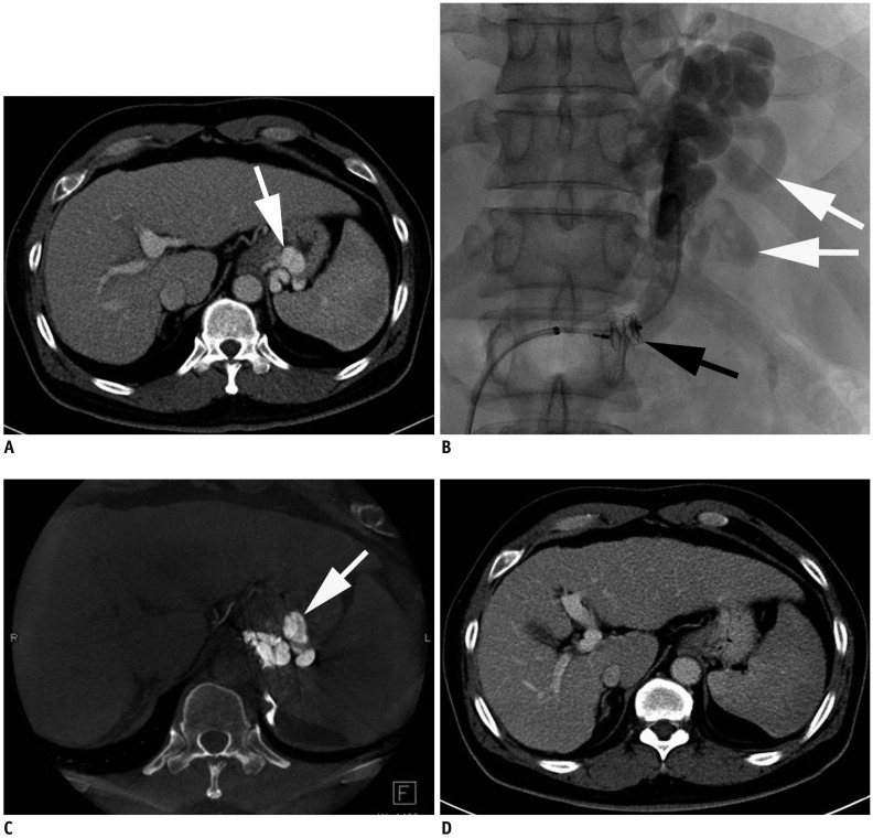
Fig. 1 52-year-old man with GV.A. Contrast-enhanced CT image obtained before PARTO shows GV (white arrow). B. After vascular plug (black arrow) placement in narrowest portion of portosystemic shunt via left adrenal vein, additional embolization of splenorenal shunt, GV, and afferent vein (white arrows) from splenic vein was performed using gelatin sponges through 4-F catheter. C. Non-contrast CBCT image obtained immediately after injection of gelatin sponges showed complete embolization of GV (white arrow). D. Contrast-enhanced CT image obtained three months after PARTO showed complete obliteration of GV. CBCT = cone-beam computed tomography, GV = gastric varices, PARTO = plug-assisted retrograde transvenous obliteration
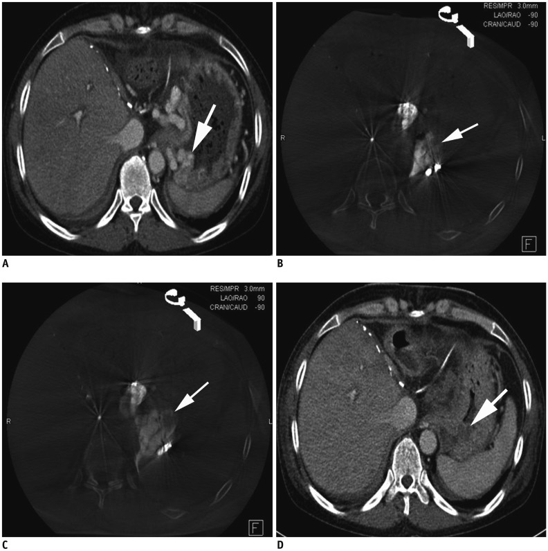
Fig. 2 48-year-old woman with GV.A. Contrast-enhanced CT image obtained before PARTO shows GV (arrow). B. Non-contrast CBCT image obtained immediately after injection of gelatin sponges showed incomplete embolization of GV (arrow). C. Second non-contrast CBCT image obtained immediately after additional injection of gelatin sponges shows complete embolization of GV (arrow). D. Contrast-enhanced CT image obtained one day after PARTO shows complete thrombosis of GV (arrow).
Reference
-
1. de Franchis R, Primignani M. Natural history of portal hypertension in patients with cirrhosis. Clin Liver Dis. 2001; 5:645–663. PMID: 11565135.
Article2. Trudeau W, Prindiville T. Endoscopic injection sclerosis in bleeding gastric varices. Gastrointest Endosc. 1986; 32:264–268. PMID: 3488937.
Article3. Sarin SK, Lahoti D, Saxena SP, Murthy NS, Makwana UK. Prevalence, classification and natural history of gastric varices: a long-term follow-up study in 568 portal hypertension patients. Hepatology. 1992; 16:1343–1349. PMID: 1446890.
Article4. Kanagawa H, Mima S, Kouyama H, Gotoh K, Uchida T, Okuda K. Treatment of gastric fundal varices by balloon-occluded retrograde transvenous obliteration. J Gastroenterol Hepatol. 1996; 11:51–58. PMID: 8672742.
Article5. Chikamori F, Kuniyoshi N, Shibuya S, Takase Y. Eight years of experience with transjugular retrograde obliteration for gastric varices with gastrorenal shunts. Surgery. 2001; 129:414–420. PMID: 11283531.
Article6. Fukuda T, Hirota S, Sugimura K. Long-term results of balloon-occluded retrograde transvenous obliteration for the treatment of gastric varices and hepatic encephalopathy. J Vasc Interv Radiol. 2001; 12:327–336. PMID: 11287510.
Article7. Cho SK, Shin SW, Lee IH, Do YS, Choo SW, Park KB, et al. Balloon-occluded retrograde transvenous obliteration of gastric varices: outcomes and complications in 49 patients. AJR Am J Roentgenol. 2007; 189:W365–W372. PMID: 18029851.
Article8. Choi SY, Won JY, Kim KA, Lee DY, Lee KH. Foam sclerotherapy using polidocanol for balloon-occluded retrograde transvenous obliteration (BRTO). Eur Radiol. 2011; 21:122–129. PMID: 20737152.
Article9. Chang IS, Park SW, Kwon SY, Choe WH, Cheon YK, Shim CS, et al. Efficacy and safety of balloon-occluded retrograde transvenous obliteration with sodium tetradecyl sulfate liquid sclerotherapy. Korean J Radiol. 2016; 17:224–229. PMID: 26957907.
Article10. Ninoi T, Nishida N, Kaminou T, Sakai Y, Kitayama T, Hamuro M, et al. Balloon-occluded retrograde transvenous obliteration of gastric varices with gastrorenal shunt: long-term follow-up in 78 patients. AJR Am J Roentgenol. 2005; 184:1340–1346. PMID: 15788621.
Article11. Kim SK, Lee KA, Sauk S, Korenblat K. Comparison of transjugular intrahepatic portosystemic shunt with covered stent and balloon-occluded retrograde transvenous obliteration in managing isolated gastric varices. Korean J Radiol. 2017; 18:345–354. PMID: 28246514.
Article12. Akahoshi T, Hashizume M, Tomikawa M, Kawanaka H, Yamaguchi S, Konishi K, et al. Long-term results of balloon-occluded retrograde transvenous obliteration for gastric variceal bleeding and risky gastric varices: a 10-year experience. J Gastroenterol Hepatol. 2008; 23:1702–1709. PMID: 18713295.
Article13. Sabri SS, Swee W, Turba UC, Saad WE, Park AW, Al-Osaimi AM, et al. Bleeding gastric varices obliteration with balloon-occluded retrograde transvenous obliteration using sodium tetradecyl sulfate foam. J Vasc Interv Radiol. 2011; 22:309–316. PMID: 21353984.
Article14. Kim MY, Um SH, Baik SK, Seo YS, Park SY, Lee JI, et al. Clinical features and outcomes of gastric variceal bleeding: retrospective Korean multicenter data. Clin Mol Hepatol. 2013; 19:36–44. PMID: 23593608.
Article15. Imai Y, Nakazawa M, Ando S, Sugawara K, Mochida S. Long-term outcome of 154 patients receiving balloon-occluded retrograde transvenous obliteration for gastric fundal varices. J Gastroenterol Hepatol. 2016; 31:1844–1850. PMID: 27003222.
Article16. Gwon DI, Ko GY, Yoon HK, Sung KB, Kim JH, Shin JH, et al. Gastric varices and hepatic encephalopathy: treatment with vascular plug and gelatin sponge-assisted retrograde transvenous obliteration--a primary report. Radiology. 2013; 268:281–287. PMID: 23481167.
Article17. Gwon DI, Kim YH, Ko GY, Kim JW, Ko HK, Kim JH, et al. Vascular plug-assisted retrograde transvenous obliteration for the treatment of gastric varices and hepatic encephalopathy: a prospective multicenter study. J Vasc Interv Radiol. 2015; 26:1589–1595. PMID: 26316136.
Article18. Kim T, Yang H, Lee CK, Kim GB. Vascular plug assisted retrograde transvenous obliteration (PARTO) for gastric varix bleeding patients in the emergent clinical setting. Yonsei Med J. 2016; 57:973–979. PMID: 27189294.
Article19. Chang MY, Kim MD, Kim T, Shin W, Shin M, Kim GM, et al. Plug-assisted retrograde transvenous obliteration for the treatment of gastric variceal hemorrhage. Korean J Radiol. 2016; 17:230–238. PMID: 26957908.
Article20. Deschamps F, Solomon SB, Thornton RH, Rao P, Hakime A, Kuoch V, et al. Computed analysis of three-dimensional cone-beam computed tomography angiography for determination of tumor-feeding vessels during chemoembolization of liver tumor: a pilot study. Cardiovasc Intervent Radiol. 2010; 33:1235–1242. PMID: 20390271.
Article21. Hirota S, Nakao N, Yamamoto S, Kobayashi K, Maeda H, Ishikura R, et al. Cone-beam CT with flat-panel-detector digital angiography system: early experience in abdominal interventional procedures. Cardiovasc Intervent Radiol. 2006; 29:1034–1038. PMID: 16988877.
Article22. Koizumi J, Hashimoto T, Myojin K, Fukushima T, Ichikawa T, Kagawa T, et al. C-arm CT-guided foam sclerotherapy for the treatment of gastric varices. J Vasc Interv Radiol. 2010; 21:1583–1587. PMID: 20801674.
Article23. Yamagami T, Yoshimatsu R, Miura H, Tanaka O, Yasui K, Yagi N, et al. Usefulness of cone-beam computed tomography during balloon-occluded retrograde transvenous obliteration. Minim Invasive Ther Allied Technol. 2013; 22:359–363. PMID: 23808369.
Article24. Lee EW, So N, Chapman R, McWilliams JP, Loh CT, Busuttil RW, et al. Usefulness of intra-procedural cone-beam computed tomography in modified balloon-occluded retrograde transvenous obliteration of gastric varices. World J Radiol. 2016; 8:390–396. PMID: 27158425.
Article
- Full Text Links
-
- Actions
-
Cited
- CITED
-
- Close
- Share
-
- Similar articles
-
- Plug-Assisted Retrograde Transvenous Obliteration for the Treatment of Gastric Varix with Both Gastrorenal and Gastrocaval Shunts: A Case Report
- Successful Treatment of Duodenal Variceal Bleeding with Coil-Assisted Retrograde Transvenous Obliteration: A Case Report
- Plug-Assisted Retrograde Transvenous Obliteration of Spontaneous Splenorenal Shunt for Refractory Hepatic Encephalopathy: Case Series
- Plug-Assisted Retrograde Transvenous Obliteration for the Treatment of Duodenal Variceal Bleeding - A Case Report and Literature Review
- Balloon Occlusion Retrograde Transvenous Obliteration of Gastric Varices in Two Non-Cirrhotic Patients with Portal Vein Thrombosis
