Restor Dent Endod.
2017 Nov;42(4):273-281. 10.5395/rde.2017.42.4.273.
Quantification of the tug-back by measuring the pulling force and micro computed tomographic evaluation
- Affiliations
-
- 1Department of Conservative Dentistry, Wonkwang University Daejeon Dental Hospital, Daejeon, Korea. profee@wku.ac.kr
- KMID: 2403068
- DOI: http://doi.org/10.5395/rde.2017.42.4.273
Abstract
OBJECTIVES
The aims of this study were to quantify tug-back by measuring the pulling force and investigate the correlation of clinical tug-back pulling force with in vitro gutta-percha (GP) cone adaptation score using micro-computed tomography (µCT).
MATERIALS AND METHODS
Twenty-eight roots from human single-rooted teeth were divided into 2 groups. In the ProTaper Next (PTN) group, root canals were prepared with PTN, and in the ProFile (PF) group, root canals were prepared using PF (n = 14). The degree of tug-back was scored after selecting taper-matched GP cones. A novel method using a spring balance was designed to quantify the tug-back by measuring the pulling force. The correlation between tug-back scores, pulling force, and percentage of the gutta-percha occupied area (pGPOA) within apical 3 mm was investigated using µCT. The data were analyzed using Pearson's correlation analysis, one-way analysis of variance (ANOVA) and Tukey's test.
RESULTS
Specimens with a strong tug-back had a mean pulling force of 1.24 N (range, 0.15-1.70 N). This study showed a positive correlation between tug-back score, pulling force, and pGPOA. However, there was no significant difference in these factors between the PTN and PF groups. Regardless of the groups, pGPOA and pulling force were significantly higher in the specimens with a higher tug-back score (p < 0.05).
CONCLUSIONS
The degree of subjective tug-back was a definitive determinant for master cone adaptation in the root canal. The use of the tug-back scoring system and pulling force allows the interpretation of subjective tug-back in a more objective and quantitative manner.
MeSH Terms
Figure
-
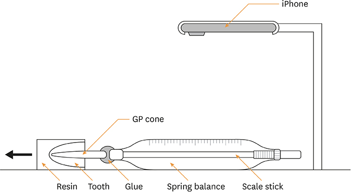
Figure 1 Schematic drawing of the device consisting of a spring balance and slow motion video to measure the pulling force (N) while removing the master cone from the root canal. GP, gutta-percha.
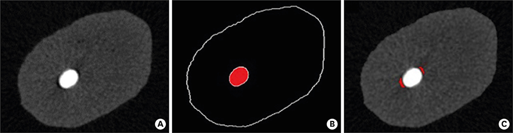
Figure 2 Example of an image analysis using image analysis software. (A) Raw image. (B) Image binarization by thresholding for GP cone, and (C) voids. GP, gutta-percha.
Reference
-
1. Inan U, Aydin C, Tunca YM, Basak F. In vitro evaluation of matched-taper single-cone obturation with a fluid filtration method. J Can Dent Assoc. 2009; 75:123.2. Gordon MP, Love RM, Chandler NP. An evaluation of .06 tapered gutta-percha cones for filling of .06 taper prepared curved root canals. Int Endod J. 2005; 38:87–96.
Article3. Schilder H. Filling root canals in three dimensions. 1967. J Endod. 2006; 32:281–290.4. Silva-Filho JM, Souza-Gabriel AE, Leoni GB, De-Bem SH, Alfredo E, Silva RG. Comparison of two techniques for selection of master gutta-percha cone using micro-computed tomography. Braz Dent J. 2013; 24:367–370.
Article5. Hembrough MW, Steiman HR, Belanger KK. Lateral condensation in canals prepared with nickel titanium rotary instruments: an evaluation of the use of three different master cones. J Endod. 2002; 28:516–519.
Article6. Eleazer P, Glickman G, McClanahan S, Webb T, Jusrman B. Glossary of endodontic terms. 8th ed. Chicago (IL): American Association of Endodontists;2012.7. Hargreaves KM, Berman LH. Cohen's pathways of the pulp expert consult. 11th ed. St. Louis (MO): Mosby;2015. p. 270–314.8. Schilder H. Vertical compaction of warm gutta-percha. In : Gerstein H, editor. Techniques in clinical endodontics. Philadelphia (PA): WB Saunders;1983. p. 76–98.9. Saatchi M, Barekatain B, Behzadian M. Comparing the apical microleakage of lateral condensation and chloroform dip techniques with a new obturation method. Dent Res J (Isfahan). 2011; 8:22–27.10. Allison DA, Michelich RJ, Walton RE. The influence of master cone adaptation on the quality of the apical seal. J Endod. 1981; 7:61–65.
Article11. Schneider SW. A comparison of canal preparations in straight and curved root canals. Oral Surg Oral Med Oral Pathol. 1971; 32:271–275.
Article12. Altman DG. Practical statistics for medical research. London: Chapman & Hall;1991. p. 396–403.13. Yoon H, Baek SH, Kum KY, Kim HC, Moon YM, Fang DY, Lee W. Fitness of gutta-percha cones in curved root canals prepared with reciprocating files correlated with tug-back sensation. J Endod. 2015; 41:102–105.
Article14. Krug R, Krastl G, Jahreis M. Technical quality of a matching-taper single-cone filling technique following rotary instrumentation compared with lateral compaction after manual preparation: a retrospective study. Clin Oral Investig. 2017; 21:643–652.
Article15. Paqué F, Ganahl D, Peters OA. Effects of root canal preparation on apical geometry assessed by micro-computed tomography. J Endod. 2009; 35:1056–1059.
Article16. Schäfer E, Köster M, Bürklein S. Percentage of gutta-percha-filled areas in canals instrumented with nickel-titanium systems and obturated with matching single cones. J Endod. 2013; 39:924–928.
Article17. Moule AJ, Kellaway R, Clarkson R, Rowell J, Macfarlane R, Lewis D, Cameron T, Atkins D. Variability of master gutta-percha cones. Aust Endod J. 2002; 28:38–43.
Article18. Lask JT, Walker MP, Kulild JC, Cunningham KP, Shull PA. Variability of the diameter and taper of size #30, 0.04 nickel-titanium rotary files. J Endod. 2006; 32:1171–1173.
Article19. Cunningham KP, Walker MP, Kulild JC, Lask JT. Variability of the diameter and taper of size #30, 0.04 gutta-percha cones. J Endod. 2006; 32:1081–1084.
Article20. Chesler MB, Tordik PA, Imamura GM, Goodell GG. Intramanufacturer diameter and taper variability of rotary instruments and their corresponding gutta-percha cones. J Endod. 2013; 39:538–541.
Article21. Mokhtari H, Rahimi S, Forough Reyhani M, Galledar S, Mokhtari Zonouzi HR. Comparison of push-out bond strength of gutta-percha to root canal dentin in single-cone and cold lateral compaction techniques with AH Plus sealer in mandibular premolars. J Dent Res Dent Clin Dent Prospect. 2015; 9:221–225.
Article22. Nagas E, Altundasar E, Serper A. The effect of master point taper on bond strength and apical sealing ability of different root canal sealers. Oral Surg Oral Med Oral Pathol Oral Radiol Endod. 2009; 107:e61–e64.
Article23. Romania C, Beltes P, Boutsioukis C, Dandakis C. Ex-vivo area-metric analysis of root canal obturation using gutta-percha cones of different taper. Int Endod J. 2009; 42:491–498.
Article24. Wu MK, Ozok AR, Wesselink PR. Sealer distribution in root canals obturated by three techniques. Int Endod J. 2000; 33:340–345.
Article25. Nica LM, Didilescu A, Rusu D, Bacila A, Stratul SI. Photomicrographic evaluation of the apical sealing capacity of three types of gutta-percha master cones: an in vitro study. Odontology. 2012; 100:54–60.
Article26. Somma F, Cretella G, Carotenuto M, Pecci R, Bedini R, De Biasi M, Angerame D. Quality of thermoplasticized and single point root fillings assessed by micro-computed tomography. Int Endod J. 2011; 44:362–369.
Article27. Ho ES, Chang JW, Cheung GS. Quality of root canal fillings using three gutta-percha obturation techniques. Restor Dent Endod. 2016; 41:22–28.
Article28. Jung M, Lommel D, Klimek J. The imaging of root canal obturation using micro-CT. Int Endod J. 2005; 38:617–626.
Article29. Simon JH. The apex: how critical is it? Gen Dent. 1994; 42:330–334.
- Full Text Links
-
- Actions
-
Cited
- CITED
-
- Close
- Share
-
- Similar articles
-
- Corrigendum: Corrections to the funding. Quantification of the tug-back by measuring the pulling force and micro computed tomographic evaluation
- Evaluation and development of digital device for measuring proximal tooth contact tightness
- Study on the Body Temperature Measuring Time and Accuracy and Reliability of Tympanic Thermometer
- Computed Tomographic Findings and Clinical Observation of Spontaneous Intracerebral Hemorrhage
- Evaluation of Patella Alignment using Computed Tomographic Image
