Zygomatic miniplates for skeletal anchorage in orthopedic correction of Class III malocclusion: A controlled clinical trial
- Affiliations
-
- 1Department of Orthodontics, Faculty of Dentistry, Gazi University, Ankara, Turkey. erdalbozkaya@gmail.com
- 2Department of Oral and Maxillofacial Surgery, Faculty of Dentistry, Gazi University, Ankara, Turkey.
- KMID: 2400389
- DOI: http://doi.org/10.4041/kjod.2017.47.2.118
Abstract
OBJECTIVE
To evaluate the effects of facemask therapy, which was anchored from the zygomatic buttresses of the maxilla by using two miniplates, in skeletal Class III patients with maxillary deficiency.
METHODS
Eighteen skeletal Class III patients (10 girls and 8 boys; mean age, 11.4 ± 1.28 years) with maxillary deficiency were treated using miniplate-anchored facemasks, and their outcomes were compared with those of a Class III control group (9 girls and 9 boys; mean age, 10.6 ± 1.12 years). Two I-shaped miniplates were placed on the right and left zygomatic buttresses of the maxilla, and a facemask was applied with a 400 g force per side. Intragroup comparisons were made using the Wilcoxon test, and intergroup comparisons were made using the Mann-Whitney U-test (p < 0.05).
RESULTS
In the treatment group, the maxilla moved 3.3 mm forward, the mandible showed posterior rotation by 1.5°, and the lower incisors were retroclined after treatment. These results were significantly different from those in the control group (p < 0.05). No significant anterior rotation of the palatal plane was observed after treatment. Moreover, changes in the sagittal positions of the maxillary incisors and molars were similar between the treatment and control groups.
CONCLUSIONS
Skeletally anchored facemask therapy is an effective method for correcting Class III malocclusions, which also minimizes the undesired dental side effects of conventional methods in the maxilla.
Keyword
Figure
-

Figure 1 A and B, Miniplates and screws used for fixation. C, Design of a miniplate.
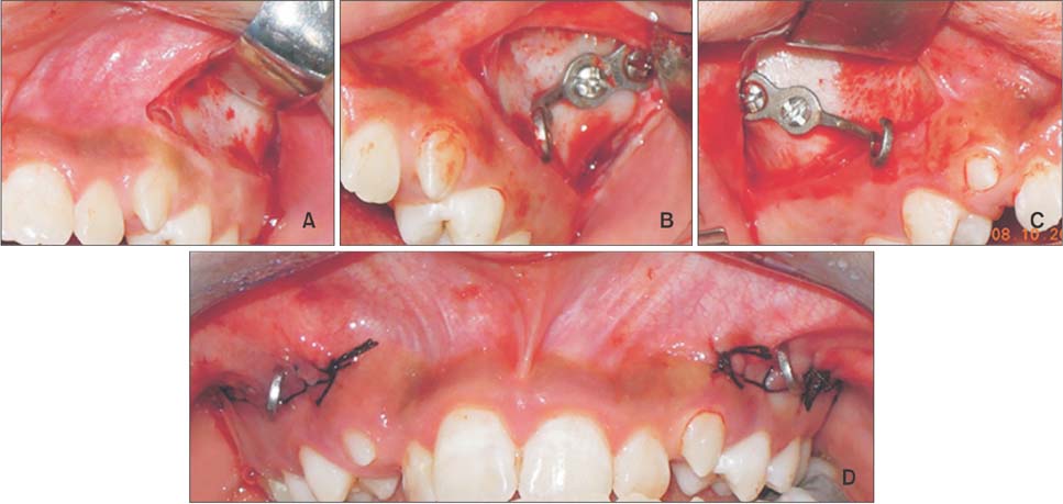
Figure 2 A, Elevated mucoperiosteal flap between the maxillary canine and the distal border of the first molar. B and C, Miniplates shaped to match the anatomic structure of the zygomatic buttresses and fixation with two self-drilling miniscrews. D, Closed flap with the C-shaped head of the miniplates exposed at the level of the soft tissue between the maxillary canine and first premolar.
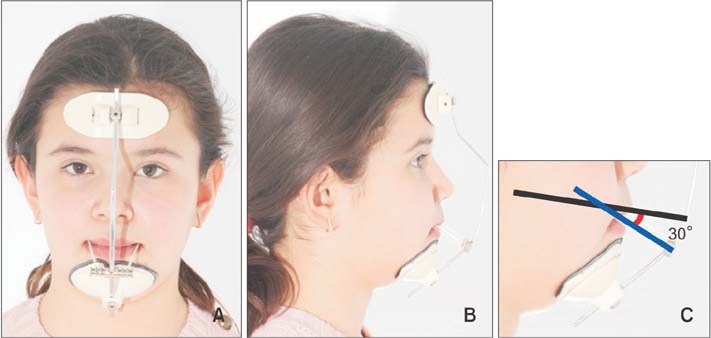
Figure 3 A and B, Facial and profile views of a patient wearing the Petit-type facemask. C, Elastics from the miniplates to the facemask (blue-colored line) extend in downward direction at an angle of 30° with reference to the occlusal plane (black-colored line).
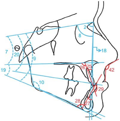
Figure 4 Cephalometric angular measurements evaluated in the present study. 2, SNA; 7, SN/ANSPNS; 8, SNB; 9, SN/GoGn; 10, ArGoMe; 18, ANB; 19, ANSPNS/GoGn; 20, SN/OP; 25, U1/NA; 26, U1/ANSPNS; 27, L1/NB; 28, L1/GoMe; 29, U1/L1; 42, nasolabial angle.

Figure 5 Cephalometric linear skeletal measurements evaluated in the present study. x, The tuberculum sella-wing (T-W) plane; y, the perpendicular plane to the T-W plane at the T point; 1, S-N; 3, Co-A; 4, A-x; 5, A-y; 6, ANS-PNS; 11, Co-Gn; 12, Go-Me; 13, Ar-Go; 14, B-x; 15, B-y; 16, Co-x; 17, Coy; 21, Wits; 22, N-ANS; 23, ANS-Me; 24, S-Go; 40, UL-S line; 41, LL-S line.
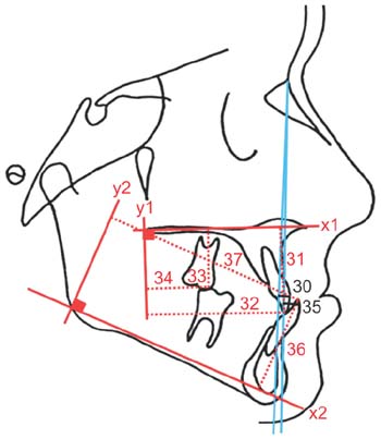
Figure 6 Cephalometric linear dental measurements evaluated in the present study. x1, The ANS-PNS plane; y1, the perpendicular plane to the ANS-PNS plane at the PNS point; x2, the Go-Gn plane; y2, the perpendicular plane to the Go-Gn plane at the Go point; 30, U1-NA; 31, U1-x1; 32, U1-y1; 33, U6-x1; 34, U6-y1; 35, L1-NB; 36, L1-x2; 37, L1-y2; 38, overjet; 39, overbite.

Figure 7 Pretreatment facial and dental images of patient. A–C, Frontal, oblique and profile view in occlusion; D–F, frontal, oblique and profile view in smiling. G, Intraoral frontal view; H, intraoral lateral view (right); I, intraoral lateral view (left); J, maxillary occlusal view; K, overjet; L, mandibular occlusal view.

Figure 8 Posttreatment facial and dental images of the same patient. A–C, Frontal, oblique and profile view in occlusion; D–F, frontal, oblique and profile view in smiling. G, Intraoral frontal view; H, intraoral lateral view (right); I, intraoral lateral view (left); J, maxillary occlusal view; K, overjet; L, mandibular occlusal view.
Reference
-
1. Tortop T, Keykubat A, Yuksel S. Facemask therapy with and without expansion. Am J Orthod Dentofacial Orthop. 2007; 132:467–474.
Article2. Gallagher RW, Miranda F, Buschang PH. Maxillary protraction: treatment and posttreatment effects. Am J Orthod Dentofacial Orthop. 1998; 113:612–619.3. Chen L, Chen R, Yang Y, Ji G, Shen G. The effects of maxillary protraction and its long-term stability--a clinical trial in Chinese adolescents. Eur J Orthod. 2012; 34:88–95.
Article4. Cha KS. Skeletal changes of maxillary protraction in patients exhibiting skeletal class III malocclusion: a comparison of three skeletal maturation groups. Angle Orthod. 2003; 73:26–35.5. Kapust AJ, Sinclair PM, Turley PK. Cephalometric effects of face mask/expansion therapy in Class III children: a comparison of three age groups. Am J Orthod Dentofacial Orthop. 1998; 113:204–212.
Article6. Kokich VG, Shapiro PA, Oswald R, Koskinen-Moffett L, Clarren SK. Ankylosed teeth as abutments for maxillary protraction: a case report. Am J Orthod. 1985; 88:303–307.
Article7. Singer SL, Henry PJ, Rosenberg I. Osseointegrated implants as an adjunct to facemask therapy: a case report. Angle Orthod. 2000; 70:253–262.8. Enacar A, Giray B, Pehlivanoglu M, Iplikcioglu H. Facemask therapy with rigid anchorage in a patient with maxillary hypoplasia and severe oligodontia. Am J Orthod Dentofacial Orthop. 2003; 123:571–577.
Article9. Hong H, Ngan P, Han G, Qi LG, Wei SH. Use of onplants as stable anchorage for facemask treatment: a case report. Angle Orthod. 2005; 75:453–460.10. De Clerck HJ, Cornelis MA, Cevidanes LH, Heymann GC, Tulloch CJ. Orthopedic traction of the maxilla with miniplates: a new perspective for treatment of midface deficiency. J Oral Maxillofac Surg. 2009; 67:2123–2129.
Article11. Kircelli BH, Pektaş ZO, Uçkan S. Orthopedic protraction with skeletal anchorage in a patient with maxillary hypoplasia and hypodontia. Angle Orthod. 2006; 76:156–163.12. Baek SH, Kim KW, Choi JY. New treatment modality for maxillary hypoplasia in cleft patients. Protraction facemask with miniplate anchorage. Angle Orthod. 2010; 80:783–791.13. Sar C, Arman-Özçırpıcı A, Uçkan S, Yazıcı AC. Comparative evaluation of maxillary protraction with or without skeletal anchorage. Am J Orthod Dentofacial Orthop. 2011; 139:636–649.
Article14. Kaya D, Kocadereli I, Kan B, Tasar F. Effects of facemask treatment anchored with miniplates after alternate rapid maxillary expansions and constrictions; a pilot study. Angle Orthod. 2011; 81:639–646.
Article15. Lee NK, Yang IH, Baek SH. The short-term treatment effects of face mask therapy in Class III patients based on the anchorage device: miniplates vs rapid maxillary expansion. Angle Orthod. 2012; 82:846–852.
Article16. Cha BK, Ngan PW. Skeletal anchorage for orthopedic correction of growing class III patients. Semin Orthod. 2011; 17:124–137.
Article17. Cha BK, Lee NK, Choi DS. Maxillary protraction treatment of skeletal Class III children using miniplate anchorage. Korean J Orthod. 2007; 37:73–84.18. Kircelli BH, Pektas ZO. Midfacial protraction with skeletally anchored face mask therapy: a novel approach and preliminary results. Am J Orthod Dentofacial Orthop. 2008; 133:440–449.
Article19. Saadia M, Torres E. Sagittal changes after maxillary protraction with expansion in class III patients in the primary, mixed, and late mixed dentitions: a longitudinal retrospective study. Am J Orthod Dentofacial Orthop. 2000; 117:669–680.
Article20. Zhou YH, Ding P, Lin Y, Qiu LX. Facemask therapy with miniplate implant anchorage in a patient with maxillary hypoplasia. Chin Med J (Engl). 2007; 120:1372–1375.
Article21. Coscia G, Addabbo F, Peluso V, D'Ambrosio E. Use of intermaxillary forces in early treatment of maxillary deficient class III patients: results of a case series. J Craniomaxillofac Surg. 2012; 40:e350–e354.
Article22. Billiet T, de Pauw G, Dermaut L. Location of the centre of resistance of the upper dentition and the nasomaxillary complex. An experimental study. Eur J Orthod. 2001; 23:263–273.
Article23. Lee HS, Choi HM, Choi DS, Jang I, Cha BK. Bone thickness of the infrazygomatic crest area in skeletal Class III growing patients: A computed tomographic study. Imaging Sci Dent. 2013; 43:261–266.
Article24. De Clerck H, Geerinckx V, Siciliano S. The zygoma anchorage system. J Clin Orthod. 2002; 36:455–459.25. Ishida T, Yoon HS, Ono T. Asymmetrical distalization of maxillary molars with zygomatic anchorage, improved superelastic nickel-titanium alloy wires, and open-coil springs. Am J Orthod Dentofacial Orthop. 2013; 144:583–593.
Article26. Arat M, Köklü A, Ozdiler E, Rübendüz M, Erdoğan B. Craniofacial growth and skeletal maturation: a mixed longitudinal study. Eur J Orthod. 2001; 23:355–361.
Article27. Melsen B, Melsen F. The postnatal development of the palatomaxillary region studied on human autopsy material. Am J Orthod. 1982; 82:329–342.
Article28. Hickham JH. Maxillary protraction therapy: diagnosis and treatment. J Clin Orthod. 1991; 25:102–113.29. Pangrazio-Kulbersh V, Berger J, Kersten G. Effects of protraction mechanics on the midface. Am J Orthod Dentofacial Orthop. 1998; 114:484–491.
Article30. Ishii H, Morita S, Takeuchi Y, Nakamura S. Treatment effect of combined maxillary protraction and chincap appliance in severe skeletal Class III cases. Am J Orthod Dentofacial Orthop. 1987; 92:304–312.
Article
- Full Text Links
-
- Actions
-
Cited
- CITED
-
- Close
- Share
-
- Similar articles
-
- Maxillary protraction using skeletal anchorage and intermaxillary elastics in Skeletal Class III patients
- Maxillary protraction treatment of skeletal Class III children using miniplate anchorage
- Class III nonsurgical treatment using indirect skeletal anchorage: A case report
- A study of the calcification of the second and the third molars in skeletal Class II and III malocclusions
- Clinical consideration of Angle's classification Class III malocclusion
