Highly Accelerated SSFP Imaging with Controlled Aliasing in Parallel Imaging and integrated-SSFP (CAIPI-iSSFP)
- Affiliations
-
- 1Department of Radiological Sciences, University of California Los Angeles, California, USA.
- 2Philips, MR Clinical Science NA, Florida, USA.
- 3Laboratory of FMRI Technology (LOFT), Stevens Neuroimaging and Informatics Institute, University of Southern California, California, USA. jwang71@gmail.com
- 4Center of Magnetic Resonance Research, University of Minnesota, Minnesota, USA.
- KMID: 2400375
- DOI: http://doi.org/10.13104/imri.2017.21.4.210
Abstract
- PURPOSE
To develop a novel combination of controlled aliasing in parallel imaging results in higher acceleration (CAIPIRINHA) with integrated SSFP (CAIPI-iSSFP) for accelerated SSFP imaging without banding artifacts at 3T.
MATERIALS AND METHODS
CAIPI-iSSFP was developed by adding a dephasing gradient to the balanced SSFP (bSSFP) pulse sequence with a gradient area that results in 2Ï€ dephasing across a single pixel. Extended phase graph (EPG) simulations were performed to show the signal behaviors of iSSFP, bSSFP, and RF-spoiled gradient echo (SPGR) sequences. In vivo experiments were performed for brain and abdominal imaging at 3T with simultaneous multi-slice (SMS) acceleration factors of 2, 3 and 4 with CAIPI-iSSFP and CAIPI-bSSFP. The image quality was evaluated by measuring the relative contrast-to-noise ratio (CNR) and by qualitatively assessing banding artifact removal in the brain.
RESULTS
Banding artifacts were removed using CAIPI-iSSFP compared to CAIPI-bSSFP up to an SMS factor of 4 and 3 on brain and liver imaging, respectively. The relative CNRs between gray and white matter were on average 18% lower in CAIPI-iSSFP compared to that of CAIPI-bSSFP.
CONCLUSION
This study demonstrated that CAIPI-iSSFP provides up to a factor of four acceleration, while minimizing the banding artifacts with up to a 20% decrease in the relative CNR.
Keyword
MeSH Terms
Figure
-
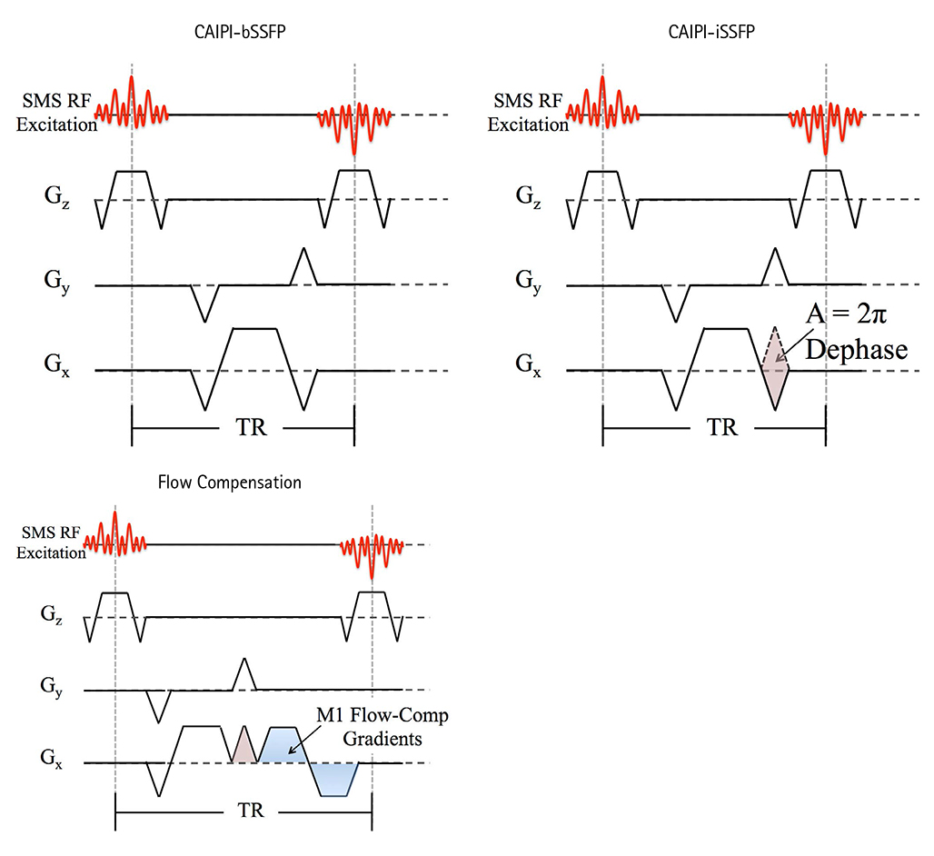
Fig. 1 Pulse sequence diagrams for CAIPI-bSSFP (left) and CAIPI-iSSFP (right) and CAIPI-iSSFP with flow compensation gradients. CAIPI-bSSFP and CAIPI-iSSFP were the same except for the added dephasing gradient in the readout direction, where it caused a 2π dephasing of the spins within a voxel, which resulted in an averaging of the bSSFP signal profile. To compensate for and reduce flow related artifacts and signal loss in CAIPI-iSSFP, bipolar gradients were added. CAIPI-bSSFP = controlled aliasing in parallel imaging and balanced steady-state free precession; CAIPI-iSSFP = controlled aliasing in parallel imaging and integrated SSFP; RF = radiofrequency; SMS = simultaneous multi-slice; TR = repetition time
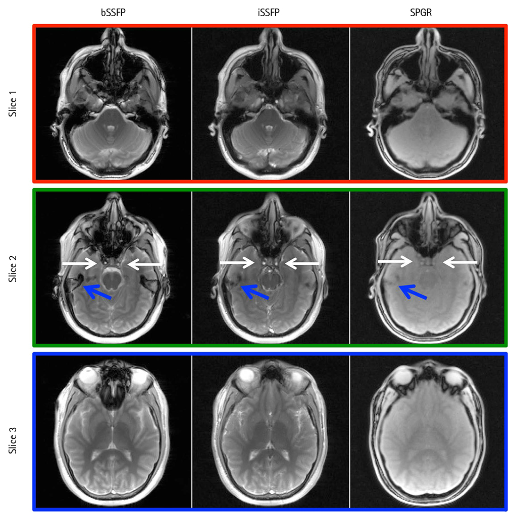
Fig. 2 Comparison of bSSFP, iSSFP, and SPGR images without CAIPIRINHA acceleration methods. The iSSFP contrast was more similar to that of bSSFP than SPGR. White arrows indicate a blood signal that was visible in bSSFP and iSSFP, but not SPGR. Blue arrows highlight the banding artifacts in bSSFP image that were not present in iSSFP and SPGR. bSSFP = balanced steady-state free precession; CAIPIRINHA = controlled aliasing in parallel imaging results in higher acceleration; iSSFP = integrated SSFP; SPGR = spoiled gradient echo
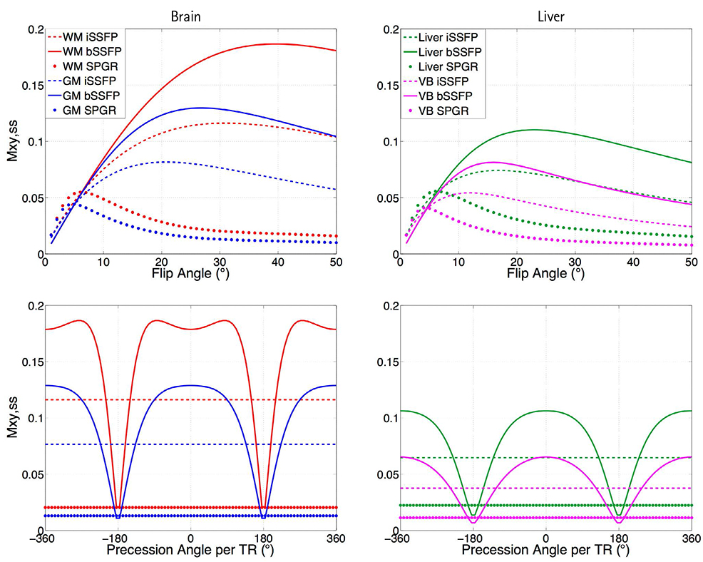
Fig. 3 Numerical simulations plotting the signal profiles of bSSFP, iSSFP, and SPGR as a function of the flip angle (top) and off resonance (bottom) for white matter (WM), gray matter (GM), liver, and venous blood (VB). The simulated signal for iSSFP was less than bSSFP and higher than SPGR for all tissue and blood, and was not sensitive to off-resonance. bSSFP = balanced steady-state free precession; iSSFP = integrated SSFP; SPGR = spoiled gradient echo; TR = repetition time
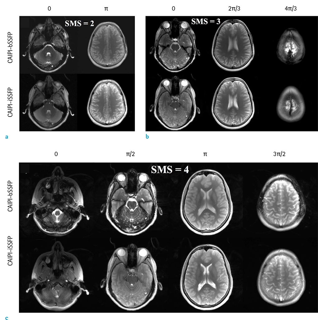
Fig. 4 Comparison of CAIPI-iSSFP and CAIPI-bSSFP with SMS acceleration factors of 2 (a), 3 (b), and 4 (c). Examples of unaliased images from a single simultaneously excited slice are shown. The phase modulation for each of the images is shown above. There were banding artifacts clearly present in b) and c) in the phase modulations of 0 and π/2 respectively, while in the CAIPI-iSSFP images the banding artifacts were not visible. The image contrast between the different tissues was comparable between the two sequences, and the noise did not significantly degrade the image quality with an SMS factor of 4. CAIPI-bSSFP = controlled aliasing in parallel imaging and balanced steady-state free precession; CAIPI-iSSFP = controlled aliasing in parallel imaging and integrated SSFP; SMS = simultaneous multi-slice
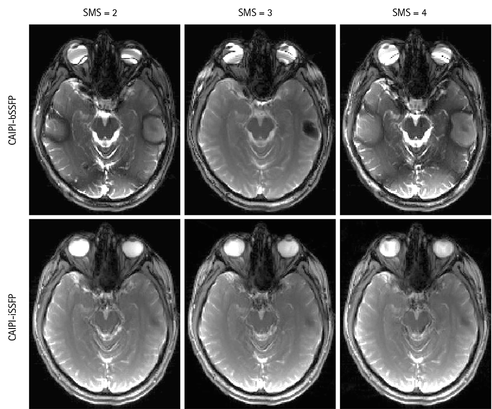
Fig. 5 Example images showing SMS factors of 2, 3, and 4 with CAIPI-iSSFP (bottom) and CAIPI-bSSFP (top) sequences using a long TR of 8.4 ms. There were severe banding artifacts in the CAIPI-bSSFP images; whereas, the CAIPI-iSSFP images did not have banding artifacts. CAIPI-bSSFP = controlled aliasing in parallel imaging and balanced steady-state free precession; CAIPI-iSSFP = controlled aliasing in parallel imaging and integrated SSFP; SMS = simultaneous multi-slice

Fig. 6 Plots showing the measured relative CNR of white matter and ventricles (a), and white and gray matter (b) for bSSFP, iSSFP, and SPGR with CAIPIRINHA acceleration SMS = 2, 3, and 4. The relative CNR was less for iSSFP compared to bSSFP, but had a much higher relative CNR than SPGR. As the SMS increased, the relative CNR decreased due to the g-factor. bSSFP = balanced steady-state free precession; CAIPIRINHA = controlled aliasing in parallel imaging results in higher acceleration; CNR = contrast-to-noise ratio; iSSFP = integrated SSFP; SMS = simultaneous multi-slice; SPGR = spoiled gradient echo

Fig. 7 CAIPI-bSSFP and CAIPI-iSSFP with SMS factor of 3 and iSSFP with flow compensation liver scan. The blue arrows identify the banding that was present in the CAIPI-bSSFP images. Spinal fluid and VB signal was suppressed in CAIPI-iSSFP due to the spoiling. However, the banding artifacts were removed and the aorta still showed a bright signal. Adding the M1 flow compensation improved the spinal fluid and VB signal (red arrows). CAIPI-bSSFP = controlled aliasing in parallel imaging and balanced steady-state free precession; CAIPI-iSSFP = controlled aliasing in parallel imaging and integrated SSFP; SMS = simultaneous multi-slice; VB = venous blood
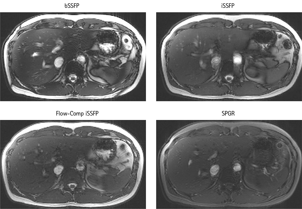
Fig. 8 Comparison of bSSFP, iSSFP, iSSFP with M1 flow compensation, and SPGR sequence images without CAIPIRINHA in the liver. The VB signal in the iSSFP and SPGR images was significantly less due to the spoiling. However, the banding artifacts were removed compared to bSSFP. The flow compensation did improve the VB ghosting artifacts; however, some of the VB signal within the liver was not as bright. bSSFP = balanced steady-state free precession; CAIPIRINHA = controlled aliasing in parallel imaging results in higher acceleration; iSSFP = integrated SSFP; VB = venous blood
Reference
-
1. Miller KL, Tijssen RH, Stikov N, Okell TW. Steady-state MRI: methods for neuroimaging. Imaging Med. 2011; 3:93–105.
Article2. Carr H. Steady-state free precession in nuclear magnetic resonance. Phys Rev. 1958; 112:1693–1701.
Article3. Bieri O, Scheffler K. Fundamentals of balanced steady state free precession MRI. J Magn Reson Imaging. 2013; 38:2–11.
Article4. Scheffler K, Lehnhardt S. Principles and applications of balanced SSFP techniques. Eur Radiol. 2003; 13:2409–2418.
Article5. Oppelt A, Graumann R, Barfuß H, Fischer H, Hartl W, Schajor W. FISP: a new fast MRI sequence. Electromedica. 1986; 54:15–18.6. Duerk JL, Lewin JS, Wendt M, Petersilge C. Remember true FISP? A high SNR, near 1-second imaging method for T2-like contrast in interventional MRI at .2 T. J Magn Reson Imaging. 1998; 8:203–220.7. Pruessmann KP, Weiger M, Scheidegger MB, Boesiger P. SENSE: sensitivity encoding for fast MRI. Magn Reson Med. 1999; 42:952–962.
Article8. Robson PM, Grant AK, Madhuranthakam AJ, Lattanzi R, Sodickson DK, McKenzie CA. Comprehensive quantification of signal-to-noise ratio and g-factor for image-based and k-space-based parallel imaging reconstructions. Magn Reson Med. 2008; 60:895–907.9. Larkman DJ, Hajnal JV, Herlihy AH, Coutts GA, Young IR, Ehnholm G. Use of multicoil arrays for separation of signal from multiple slices simultaneously excited. J Magn Reson Imaging. 2001; 13:313–317.
Article10. Setsompop K, Cohen-Adad J, Gagoski BA, et al. Improving diffusion MRI using simultaneous multi-slice echo planar imaging. Neuroimage. 2012; 63:569–580.
Article11. Breuer FA, Blaimer M, Heidemann RM, Mueller MF, Griswold MA, Jakob PM. Controlled aliasing in parallel imaging results in higher acceleration (CAIPIRINHA) for multi-slice imaging. Magn Reson Med. 2005; 53:684–691.
Article12. Stäb D, Ritter CO, Breuer FA, Weng AM, Hahn D, Köstler H. CAIPIRINHA accelerated SSFP imaging. Magn Reson Med. 2011; 65:157–164.
Article13. Haacke EM, Wielopolski PA, Tkach JA, Modic MT. Steady-state free precession imaging in the presence of motion: application for improved visualization of the cerebrospinal fluid. Radiology. 1990; 175:545–552.
Article14. Hargreaves BA. Rapid gradient-echo imaging. J Magn Reson Imaging. 2012; 36:1300–1313.
Article15. Khajehim M, Nasiraei-Moghaddam A, Hossein-Zadeh GA, Martin T, Wang D. A quantitative analysis of fMRI induced phase changes using averaged-BOSS (A-BOSS). In : In Proceedings of the 23rd Scientific Meeting of International Society for Magnetic Resonance in Medicine; Toronto. 2015. p. 3921.16. Shams Z, Moghaddam AN. Averaged-BOSS: feasibility study and preliminary results. In : In Proceedings of the 22nd Scientific Meeting of International Society for Magnetic Resonance in Medicine; Milan. 2014. p. 4216.17. Zur Y, Wood ML, Neuringer LJ. Spoiling of transverse magnetization in steady-state sequences. Magn Reson Med. 1991; 21:251–263.
Article18. Crawley AP, Wood ML, Henkelman RM. Elimination of transverse coherences in FLASH MRI. Magn Reson Med. 1988; 8:248–260.
Article19. Sekihara K. Steady-state magnetizations in rapid NMR imaging using small flip angles and short repetition intervals. IEEE Trans Med Imaging. 1987; 6:157–164.
Article20. Scheffler IE, Elson EL, Baldwin RL. Helix formation by dAT oligomers. I. Hairpin and straight-chain helices. J Mol Biol. 1968; 36:291–304.21. Breuer FA, Blaimer M, Mueller MF, et al. Controlled aliasing in volumetric parallel imaging (2D CAIPIRINHA). Magn Reson Med. 2006; 55:549–556.
Article22. Haacke EM, Lenz GW. Improving MR image quality in the presence of motion by using rephasing gradients. AJR Am J Roentgenol. 1987; 148:1251–1258.
Article23. Axel L, Morton D. MR flow imaging by velocity-compensated/uncompensated difference images. J Comput Assist Tomogr. 1987; 11:31–34.
Article24. Simonetti OP, Wendt RE 3rd, Duerk JL. Significance of the point of expansion in interpretation of gradient moments and motion sensitivity. J Magn Reson Imaging. 1991; 1:569–577.
Article25. Weigel M. Extended phase graphs: dephasing, RF pulses, and echoes - pure and simple. J Magn Reson Imaging. 2015; 41:266–295.
Article26. Wansapura JP, Holland SK, Dunn RS, Ball WS Jr. NMR relaxation times in the human brain at 3.0 tesla. J Magn Reson Imaging. 1999; 9:531–553.
Article27. Wang Y, Moeller S, Li X, et al. Simultaneous multi-slice Turbo-FLASH imaging with CAIPIRINHA for whole brain distortion-free pseudo-continuous arterial spin labeling at 3 and 7 T. Neuroimage. 2015; 113:279–288.28. Xiang QS, Hoff MN. Banding artifact removal for bSSFP imaging with an elliptical signal model. Magn Reson Med. 2014; 71:927–933.
Article29. Mikaiel S, Martin T, Sung K, Wu H. Real-time golden angle radial iSSFP for interventional MRI. In : In Proceedings of the 24th Scientific Meeting of International Society for Magnetic Resonance in Medicine; Singapore. 2016. p. 3579.30. Sun K, Xue R, Zhang P, et al. Integrated SSFP for functional brain mapping at 7T with reduced susceptibility artifact. J Magn Reson. 2017; 276:22–30.31. Kim MO, Hong T, Kim DH. Multislice CAIPIRINHA using view angle tilting technique (CAIPIVAT). Tomography. 2016; 2:43–48.
Article32. Kim D, Seo H, Oh C, Han Y, Park H. Multi-slice imAGe generation using intra-slice paraLLel imaging and Inter-slice shifting (MAGGULLI). Phys Med Biol. 2016; 61:1692–1704.
Article33. Bilgic B, Gagoski BA, Cauley SF, et al. Wave-CAIPI for highly accelerated 3D imaging. Magn Reson Med. 2015; 73:2152–2162.
Article34. Setsompop K, Gagoski BA, Polimeni JR, Witzel T, Wedeen VJ, Wald LL. Blipped-controlled aliasing in parallel imaging for simultaneous multislice echo planar imaging with reduced g-factor penalty. Magn Reson Med. 2012; 67:1210–1224.
Article35. Bieri O, Scheffler K. Flow compensation in balanced SSFP sequences. Magn Reson Med. 2005; 54:901–907.
Article36. Wang Y, Shao X, Martin T, Moeller S, Yacoub E, Wang DJ. Phase-cycled simultaneous multislice balanced SSFP imaging with CAIPIRINHA for efficient banding reduction. Magn Reson Med. 2016; 76:1764–1774.
Article37. Gagoski BA, Bilgic B, Eichner C, et al. RARE/turbo spin echo imaging with simultaneous multislice wave-CAIPI. Magn Reson Med. 2015; 73:929–938.
Article
- Full Text Links
-
- Actions
-
Cited
- CITED
-
- Close
- Share
-
- Similar articles
-
- Feasibility of Three-Dimensional Balanced Steady-State Free Precession Cine Magnetic Resonance Imaging Combined with an Image Denoising Technique to Evaluate Cardiac Function in Children with Repaired Tetralogy of Fallot
- In Vivo and In Vitro Studies of the Steady State Free Precession-Diffusion-Weighted MR Imagings on Low b-value: Validation and Application to Bone Marrow Pathology
- Diagnostic Performance of Diffusion-Weighted Steady-State Free Precession in Differential Diagnosis of Neoplastic and Benign Osteoporotic Vertebral Compression Fractures: Comparison to Diffusion-Weighted Echo-Planar Imaging
- 3D Whole-Heart Coronary MR Angiography at 1.5T in Healthy Volunteers: Comparison between Unenhanced SSFP and Gd-Enhanced FLASH Sequences
- The Usefulness of Stress Perfusion MR using Steady State Free Precession Sequence for Depiction of Significant Coronary Artery Stenosis
