Neuroanatomical Mechanism of Cerebellar Mutism After Stroke
- Affiliations
-
- 1Department of Physical Medicine and Rehabilitation, Korea University Anam Hospital, Korea University College of Medicine, Seoul, Korea. rmpyun@korea.ac.kr
- 2Department of Biomedical Sciences, Korea University Anam Hospital, Korea University College of Medicine, Seoul, Korea.
- 3Department of Rehabilitation Medicine, Bundang Jesaeng General Hospital, Seongnam, Korea.
- 4Brain Convergence Research Center, Korea University Anam Hospital, Korea University College of Medicine, Seoul, Korea.
- KMID: 2400293
- DOI: http://doi.org/10.5535/arm.2017.41.6.1076
Abstract
- Cerebellar mutism (CM) is a rare neurological condition characterized by lack of speech due to cerebellar lesions. CM is often reported in children. We describe a rare case of CM after spontaneous cerebellar hemorrhage. The patient showed mutism, irritability, decreased spontaneous movements and oropharyngeal apraxia. Diffusion tensor imaging revealed significant volume reduction of medial frontal projection fibers from the corpus callosum. In Tracts Constrained by UnderLying Anatomy (TRACULA) analysis, forceps major and minor and bilateral cingulum-angular bundles were not visualized. Cerebello-frontal pathway reconstructed from the FMRIB Software Library showed continuity of fibers, with decreased number of fibers on qualitative analysis. These results suggest that cerebello-frontal disconnection may be a neuroanatomical mechanism of CM. Damage of brain network between occipital lobe, cingulate and cerebellum caused by hemorrhage may also have role in the mechanism of CM in our case.
MeSH Terms
Figure
-
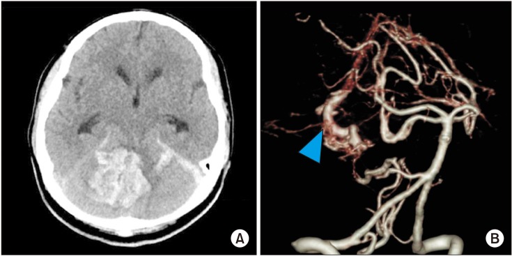
Fig. 1 Computed tomography scan showing a large amount of cerebellar hemorrhage (A) and tangled vascular structure (arrowhead) (B).

Fig. 2 Corticospinal tract (A), arcuate fasciculus (B), axial view (C) and sagittal view (D) of the corpus callosum with significant fiber loss projecting on medial frontal cortex (arrow).

Fig. 3 All tracts reconstructed by TRACULA (Tracts Constrained by UnderLying Anatomy). Forceps major, forceps minor (arrowhead) (A-a), and bilateral cingulum-angular bundles (arrow) (A-b) of a healthy control were not visualized in our case (B-a, B-b).
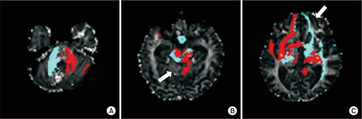
Fig. 4 Fronto-cerebellar pathway reconstructed by the FMRIB Software Library (FSL). (A) At the cerebellar hemorrhage level. (B) Decreased fronto-cerebellar fibers (arrows) between the right cerebellum and left frontal cortex was seen at the caudal midbrain level of cerebellar decussation and (C) prefrontal area.
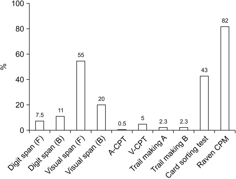
Fig. 5 Results of cognitive evaluation at 15 months after stroke. Attention and executive function scores were decreased in our case. A-CPT, auditory continuous performance test; V-CPT, visual continuous performance test; Raven CPM, Raven Coloured Progressive Matrices.
Reference
-
1. Kuper M, Timmann D. Cerebellar mutism. Brain Lang. 2013; 127:327–333. PMID: 23398780.2. Catsman-Berrevoets CE, Aarsen FK. The spectrum of neurobehavioural deficits in the Posterior Fossa Syndrome in children after cerebellar tumour surgery. Cortex. 2010; 46:933–946. PMID: 20116053.
Article3. Dubey A, Sung WS, Shaya M, Patwardhan R, Willis B, Smith D, et al. Complications of posterior cranial fossa surgery: an institutional experience of 500 patients. Surg Neurol. 2009; 72:369–375. PMID: 19604553.4. Nagaratnam N, Nagaratnam K, Ng K, Diu P. Akinetic mutism following stroke. J Clin Neurosci. 2004; 11:25–30. PMID: 14642361.
Article5. Marien P, Verslegers L, Moens M, Dua G, Herregods P, Verhoeven J. Posterior fossa syndrome after cerebellar stroke. Cerebellum. 2013; 12:686–691. PMID: 23575947.
Article6. Soelva V, Hernaiz Driever P, Abbushi A, Rueckriegel S, Bruhn H, Eisner W, et al. Fronto-cerebellar fiber tractography in pediatric patients following posterior fossa tumor surgery. Childs Nerv Syst. 2013; 29:597–607. PMID: 23184224.
Article7. Law N, Greenberg M, Bouffet E, Taylor MD, Laughlin S, Strother D, et al. Clinical and neuroanatomical predictors of cerebellar mutism syndrome. Neuro Oncol. 2012; 14:1294–1303. PMID: 22952198.
Article8. Assaf Y, Ben-Sira L, Constantini S, Chang LC, Beni-Adani L. Diffusion tensor imaging in hydrocephalus: initial experience. AJNR Am J Neuroradiol. 2006; 27:1717–1724. PMID: 16971621.
- Full Text Links
-
- Actions
-
Cited
- CITED
-
- Close
- Share
-
- Similar articles
-
- Transient Cerebellar Mutism after Total Removal of Medulloblastoma in a Child: Case Report
- Transient Mutism following Resection of 4th Ventricle Choroid Plexus Papilloma in a 6-Year-Old Girl
- Remote Cerebellar Hemorrhage Presenting with Cerebellar Mutism after Spinal Surgery: An Unusual Case Report
- Mutism after Posterior Fossa Tumor Surgery in a Child: Case Report
- Akinetic Mutism and Cognitive-Affective Syndrome Caused by Unilateral PICA Infarction
