Conservative management of a dentigerous cyst associated with eruption of teeth in a 7-year-old girl: a case report
- Affiliations
-
- 1Department of Oral and Maxillofacial Surgery, Faculty of Dentistry, İzmir Katip Çelebi University, İzmir, Turkey. onursahin43@hotmail.com
- KMID: 2399917
- DOI: http://doi.org/10.5125/jkaoms.2017.43.S1.S1
Abstract
- Dentigerous cysts are benign odontogenic cysts that are related to the crowns of permanent teeth. The lesion in this study was detected in a routine panoramic radiograph that revealed a well-defined osteolytic lesion that measured 2.5 cm in diameter, with the crown of the mandibular permanent second premolar displaced to the lower border of the mandible. The apex of the tooth was still open. The aim of this article was to report the case of a 7-year-old girl with a dentigerous cyst associated with the tooth buds of premolars. The therapeutic approach consisted of extraction of the primary molar and marsupialization of the lesion. After 40 months of follow-up, spontaneous eruption of the impacted premolar was observed. In conclusion, marsupialization can be the first treatment choice for conservative management of dentigerous cysts in pre-adolescents.
MeSH Terms
Figure
-
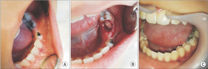
Fig. 1 Intraoral photographs. A. Intraoral view showing buccal expansion in the region of the primary mandibular left second molar. B. Decompression of the cyst was performed using a silicone tube through the socket of the tooth extraction over the lesion. C. Postoperative view of the second premolar, which erupted after 46 months.
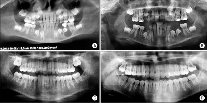
Fig. 2 Panoramic radiographs. A. Radiograph view demonstrating the dentigerous cyst related to the unerupted second premolar. B. Postoperative radiographic view after 9 months, displaying the decreased radiolucency around the second premolar. C. Postoperative 40 months. D. Postoperative 46 months.
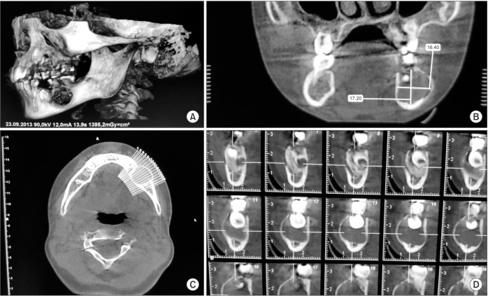
Fig. 3 A. Cone-beam computed tomography image of the lesion shows a well-defined lesion in the mandibular left region surronding the crown of the unerupted second premolar. B. Coronal view. C. Axial view. D. Sagittal view.
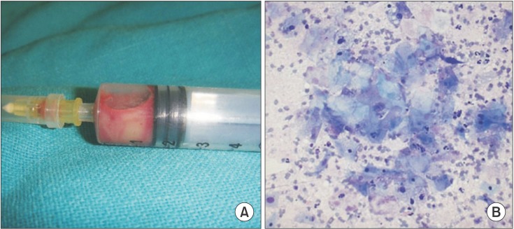
Fig. 4 A. Photograph of the aspirated fluid. B. The cytopathologic analysis of the aspirate showing a mucoid material, clumps of benign epithelial cells, and a large amount of cyst macrophages (Diff-Quick staining, ×200).
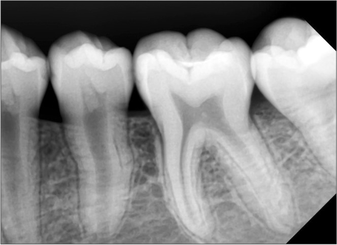
Fig. 5 Periapical radiograph of the second premolar at postoperative 46 months. Root formation of the mandibular left second premolar was almost complete.
Reference
-
1. Ikeshima A, Tamura Y. Differential diagnosis between dentigerous cyst and benign tumor with an embedded tooth. J Oral Sci. 2002; 44:13–17. PMID: 12058865.
Article2. Lustig JP, Schwartz-Arad D, Shapira A. Odontogenic cysts related to pulpotomized deciduous molars: clinical features and treatment outcome. Oral Surg Oral Med Oral Pathol Oral Radiol Endod. 1999; 87:499–503. PMID: 10225634.3. Muthray E, Desai J, Suleman Y, Meer S. Inflammatory dentigerous cyst in a 3 year old South African black male: a case report. SADJ. 2006; 61:252254–255. PMID: 16977954.4. Bodner L, Woldenberg Y, Bar-Ziv J. Radiographic features of large cystic lesions of the jaws in children. Pediatr Radiol. 2003; 33:3–6. PMID: 12497227.
Article5. Moro Antonio JM, Puente M. Surgical-orthodontic treatment of an impacted canine with a dentigerous cyst. J Clin Orthod. 2001; 35:491–493. PMID: 11589095.6. Sun KT, Chen MY, Chiang HH, Tsai HH. Treatment of large jaw bone cysts in children. J Dent Child (Chic). 2009; 76:217–222. PMID: 19941764.7. Wong M. Surgical fenestration of large periapical lesions. J Endod. 1991; 17:516–521. PMID: 1812199.
Article8. Weber AL. Imaging of cysts and odontogenic tumors of the jaw. Definition and classification. Radiol Clin North Am. 1993; 31:101–120. PMID: 8419968.9. Ertas U, Yavuz MS. Interesting eruption of 4 teeth associated with a large dentigerous cyst in mandible by only marsupialization. J Oral Maxillofac Surg. 2003; 61:728–730. PMID: 12796888.
Article10. Stoelinga PJ. Excision of the overlying, attached mucosa, in conjunction with cyst enucleation and treatment of the bony defect with carnoy solution. Oral Maxillofac Surg Clin North Am. 2003; 15:407–414. PMID: 18088692.
Article11. Anavi Y, Gal G, Miron H, Calderon S, Allon DM. Decompression of odontogenic cystic lesions: clinical long-term study of 73 cases. Oral Surg Oral Med Oral Pathol Oral Radiol Endod. 2011; 112:164–169. PMID: 21194990.
Article12. Serra e Silva FM, Sawazaki R, de Moraes M. Eruption of teeth associated with a dentigerous cyst by only marsupialization treatment: a case report. J Dent Child (Chic). 2007; 74:228–230. PMID: 18482520.13. Hayasaki H, Ishibashi M, Nakamura S, Fukumoto S, Nonaka K. Dentigerous cyst in primary dentition: case report of a 4-year-old girl. Pediatr Dent. 2009; 31:294–297. PMID: 19722437.
- Full Text Links
-
- Actions
-
Cited
- CITED
-
- Close
- Share
-
- Similar articles
-
- Conservative treatment of dentigerous cysts; 5 cases
- Surgical and Orthodonic Treatment of Impacted Teeth Associated with Dentigerous Cysts : Case Report
- Eruption and Autotransplantation of a Permanent Tooth Related to Dentigerous Cyst in Mixed Dentition
- Normal eruption guidance of unerupted permanent teeth associated with dentigerous cyst by decompression: 5 cases report
- Forced Eruption of Severe Angulated and Impacted Permanent Teeth after Marsupialization of Dentigerous Cyst: Case Report
