Age-related trends of lesser pelvic architecture in females and males: a computed tomography pelvimetry study
- Affiliations
-
- 1Joint Laboratory of Clinical Immunology and Immunogenetics, Riga Stradins University, Riga, Latvia. oksana-kolesova@inbox.lv
- 2Department of Psychology, University of Latvia, Riga, Latvia.
- 3Institute of Anatomy and Anthropology, Riga Stradins University, Riga, Latvia.
- KMID: 2399895
- DOI: http://doi.org/10.5115/acb.2017.50.4.265
Abstract
- The pelvis and the spine form a system balancing human skeleton. Within this system, the pelvis adapts to age-related changes in the spine. Previous studies were predominantly focused on changes of pelvic parameters in the sagittal plane. The aim of this study was to reveal age-related changes of lesser pelvic dimensions at different levels of the pelvic cavity in the sagittal and coronal planes and to explore sexual dimorphism in age-related tendencies. The computed tomography pelvimetry was performed on the three-dimensional workstation. The research sample included 211 females aged 18 to 84 years and 181 males aged 18 to 82 years, who underwent an examination at the Riga East University Hospital, Clinical Center "Gailezers," Latvia. Three pelvic angles and transverse and sagittal diameters of the lesser pelvis were measured at four levels: the inlet, two axial planes in the mid-cavity, and the outlet. The results demonstrated that more pronounced age-related changes occurred in the inlet and the outlet of the lesser pelvis. The mid-cavity was less changing. The transverse diameter between acetabular centers and the sagittal diameter at the level of ischial spines were independent of age. In general, the common age-related trends were observed for pelvic parameters in females and males. A single exception was the proportion of diameters at the level of ischial spines, which decreased in males only. For parameters associated with pelvic floor diseases, age-related changes occurred in the direction of pathology.
Keyword
MeSH Terms
Figure
-
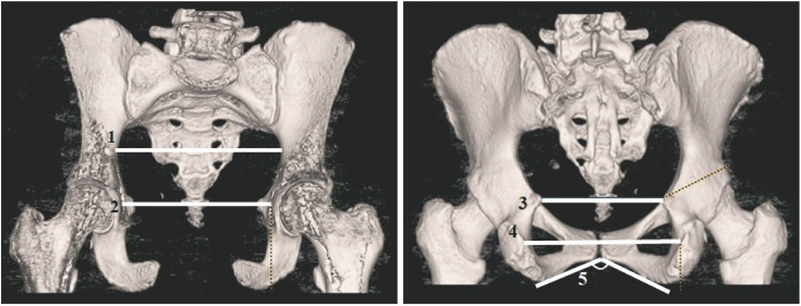
Fig. 1 3D reformatted computed tomography images: 1, transverse diameter of inlet; 2, transverse diameter of midplane 1 (biacetabular diameter); 3, transverse diameter of midplane 2 (bispinous diameter); 4, transverse diameter of outlet (bituberous diameter); 5, subpubic angle.
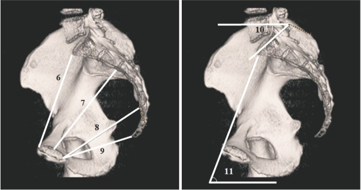
Fig. 2 3D reformatted computed tomography images: 6, sagittal diameter of inlet; 7, sagittal diameter of midplane 1; 8, sagittal diameter of midplane 2; 9, sagittal diameter of outlet; 10, sacral slope; 11, pelvic inclination.
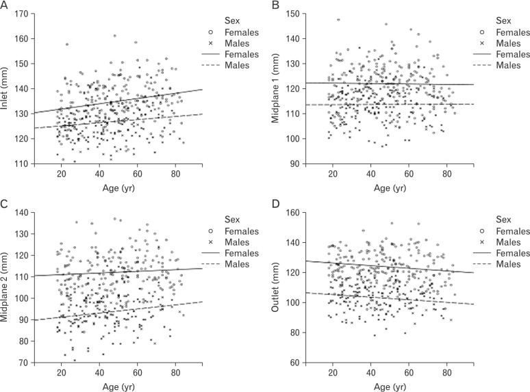
Fig. 3 Age-related trend lines for transverse diameters of the lesser pelvis in females and males. (A) Transverse diameter of inlet. (B) Transverse diameter of midplane 1. (C) Transverse diameter of midplane 2. (D) Transverse diameter of outlet.

Fig. 4 Age-related trend lines for sagittal diameters of the lesser pelvis in females and males. (A) Sagittal diameter of inlet. (B) Sagittal diameter of midplane 1. (C) Sagittal diameter of midplane 2. (D) Sagittal diameter of outlet.
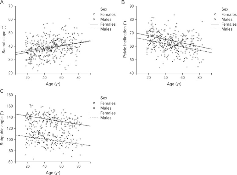
Fig. 5 Age-related trend lines for pelvic angles in females and males. (A) Sacral slope. (B) Pelvic inclination. (C) Subpubic angle.

Fig. 6 Age-related trend lines for pelvic indexes. (A) Index of inlet. (B) Index of midplane 1. (C) Index of midplane 2. (D) Index of outlet.
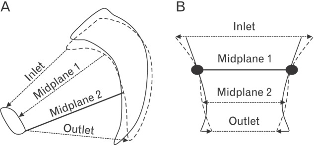
Fig. 7 Age-related changes of pelvic measures in the sagittal (A) and coronal (B) planes (dashed lines show a changing position of pelvic bones with age, while dotted lines with arrows indicate changing diameters).
Reference
-
1. Pauwels F. Beitrag zur Klärung der Beanspruchung des Beckens, insbesondere der Beckenfugen. Z Anat Entwicklungsgesch. 1948; 114:167–180. PMID: 18884221.2. Weisl H. The movements of the sacroiliac joint. Acta Anat (Basel). 1955; 23:80–91. PMID: 14349535.3. Russell JG. Moulding of the pelvic outlet. J Obstet Gynaecol Br Commonw. 1969; 76:817–820. PMID: 5823681.4. Lovejoy CO. Evolution of human walking. Sci Am. 1988; 259:118–125.5. Van Emmerik RE, McDermott WJ, Haddad JM, Van Wegen EE. Age-related changes in upper body adaptation to walking speed in human locomotion. Gait Posture. 2005; 22:233–239. PMID: 16214663.6. Moes NC. Variation in sitting pressure distribution and location of the points of maximum pressure with rotation of the pelvis, gender and body characteristics. Ergonomics. 2007; 50:536–561. PMID: 17575714.7. Kapandji A. The physiology of the joints. 6th ed. Vol. 3. Edinburg: Elsevier Limited;2008. p. 60–63. The spinal column, pelvic girdle and head.8. Schwab F, Lafage V, Boyce R, Skalli W, Farcy JP. Gravity line analysis in adult volunteers: age-related correlation with spinal parameters, pelvic parameters, and foot position. Spine (Phila Pa 1976). 2006; 31:E959–E967. PMID: 17139212.9. Amonoo-Kuofi HS. Changes in the lumbosacral angle, sacral inclination and the curvature of the lumbar spine during aging. Acta Anat (Basel). 1992; 145:373–377. PMID: 10457779.10. Peleg S, Dar G, Medlej B, Steinberg N, Masharawi Y, Latimer B, Jellema L, Peled N, Arensburg B, Hershkovitz I. Orientation of the human sacrum: anthropological perspectives and methodological approaches. Am J Phys Anthropol. 2007; 133:967–977. PMID: 17427928.11. Lazarevski MB. Pelvic bone system changes and pathogenesis of genital prolapse: a radiopelvimetric study. Gynaecol Perinatol. 2004; 13:1–12.12. Kampen WU, Tillmann B. Age-related changes in the articular cartilage of human sacroiliac joint. Anat Embryol (Berl). 1998; 198:505–513. PMID: 9833689.13. Dar G, Peleg S, Masharawi Y, Steinberg N, Rothschild BM, Peled N, Hershkovitz I. Sacroiliac joint bridging: demographical and anatomical aspects. Spine (Phila Pa 1976). 2005; 30:E429–E432. PMID: 16094261.14. Abitbol MM. Obstetrics and posture in pelvic anatomy. J Hum Evol. 1987; 16:243–255.15. Schultz AH. Sex differences in the pelves of primates. Am J Phys Anthropol. 1949; 7:401–423. PMID: 15392785.16. Tague RG. Variation in pelvic size between males and females. Am J Phys Anthropol. 1989; 80:59–71. PMID: 2801906.17. Damasceno LH, Catarin SR, Campos AD, Defino HL. Lumbar lordosis: a study of angle values and of vertebral bodies and intervertebral discs role. Acta Ortop Bras. 2006; 14:193–198.18. Mac-Thiong JM, Roussouly P, Berthonnaud E, Guigui P. Age- and sex-related variations in sagittal sacropelvic morphology and balance in asymptomatic adults. Eur Spine J. 2011; 20(Suppl 5):572–577.19. MacLennan AH, Taylor AW, Wilson DH, Wilson D. The prevalence of pelvic floor disorders and their relationship to gender, age, parity and mode of delivery. BJOG. 2000; 107:1460–1470. PMID: 11192101.20. Stav K, Alcalay M, Peleg S, Lindner A, Gayer G, Hershkovitz I. Pelvis architecture and urinary incontinence in women. Eur Urol. 2007; 52:239–244. PMID: 17207915.21. Mallett VT, Bump RC. The epidemiology of female pelvic floor dysfunction. Curr Opin Obstet Gynecol. 1994; 6:308–312. PMID: 7742490.22. DeLancey JO, Kearney R, Chou Q, Speights S, Binno S. The appearance of levator ani muscle abnormalities in magnetic resonance images after vaginal delivery. Obstet Gynecol. 2003; 101:46–53. PMID: 12517644.23. Sze EH, Kohli N, Miklos JR, Roat T, Karram MM. Computed tomography comparison of bony pelvis dimensions between women with and without genital prolapse. Obstet Gynecol. 1999; 93:229–232. PMID: 9932561.24. Nguyen JK, Lind LR, Choe JY, McKindsey F, Sinow R, Bhatia NN. Lumbosacral spine and pelvic inlet changes associated with pelvic organ prolapse. Obstet Gynecol. 2000; 95:332–336. PMID: 10711538.25. Handa VL, Pannu HK, Siddique S, Gutman R, VanRooyen J, Cundiff G. Architectural differences in the bony pelvis of women with and without pelvic floor disorders. Obstet Gynecol. 2003; 102:1283–1290. PMID: 14662216.26. Xu HN, Xia ZJ, Li BX, Yin YT, Wang F, Hu Q, Zhao Y. Investigation of correlation between diameters of pelvic inlet and outlet planes and female pelvic floor dysfunction. Eur J Obstet Gynecol Reprod Biol. 2011; 159:461–464. PMID: 21840111.27. Tabachnick BG, Fidell LS. Using multivariate statistics. 5th ed. Boston, MA: Allyn and Bacon;2007.28. Young M, Ince JG. A radiographic comparison of the male and female pelvis. J Anat. 1940; 74(Pt 3):374–385. PMID: 17104821.29. Tague RG. Do big females have big pelves? Am J Phys Anthropol. 2000; 112:377–393. PMID: 10861354.
- Full Text Links
-
- Actions
-
Cited
- CITED
-
- Close
- Share
-
- Similar articles
-
- Computed tomography based cross-sectional anatomy of the pelvis predicts surgical outcome after rectal cancer surgery
- Compare the architectural differences in the bony pelvis of Korean women and their association with the mode of delivery by computed tomography
- Three-dimensional linear and volumetric computed tomography analysis of the frontal sinus
- A study on correlation of roentgen pelvimetry and delivery mode in Korean primigravida
- Roentgenological analysis in pelvimetry in Korean women
