J Clin Neurol.
2018 Jan;14(1):100-101. 10.3988/jcn.2018.14.1.100.
Myoclonic Status Epilepticus of Unknown Etiology in an Elderly Patient
- Affiliations
-
- 1Department of Neurology, National Medical Center, Seoul, Korea. crespin97@gmail.com
- KMID: 2399606
- DOI: http://doi.org/10.3988/jcn.2018.14.1.100
Abstract
- No abstract available.
MeSH Terms
Figure
-
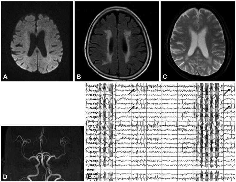
Fig. 1 Diffusion-weighted imaging (A) revealed no acute lesion and FLAIR imaging (B) revealed bilateral ischemic lesions in the subcortical areas. Gradient echo imaging (C) and MRA (D) revealed no specific abnormality. EEG showed abundant bursts of high-voltage generalized polyspikes with a normal background. Some isolated interictal spikes (arrows) were evident between or preceding the bursts (E). EEG: electroencephalogram, FLAIR: fluid-attenuated inversion recovery, MRA: magnetic resonance angiography.
Reference
-
1. Panayiotopoulos CP. A clinical guide to epileptic syndromes and their treatment. 2nd ed. London: Springer;2007. p. 63–64.2. Baysal Kirac L, Aydogdu I, Acarer A, Alpaydin S, Bayam FE, Onbasi H, et al. Myoclonic status epilepticus in six patients without epilepsy. Epilepsy Behav Case Rep. 2012; 1:10–13.
Article3. Bladin CF, Alexandrov AV, Bellavance A, Bornstein N, Chambers B, Coté R, et al. Seizures after stroke: a prospective multicenter study. Arch Neurol. 2000; 57:1617–1622.4. Schreiner A, Pohlmann-Eden B, Schwartz A, Hennerici M. Epileptic seizures in subcortical vascular encephalopathy. J Neurol Sci. 1995; 130:171–177.
Article5. De Reuck J, Nagy E, Van Maele G. Seizures and epilepsy in patients with lacunar strokes. J Neurol Sci. 2007; 263:75–78.
Article
- Full Text Links
-
- Actions
-
Cited
- CITED
-
- Close
- Share
-
- Similar articles
-
- Is the Intensive Anticonvulsant Treatment for Control of Acute Posthypoxic Myoclonic Status Epilepticus Necessary?
- A Case of Severe Myoclonic Epilepsy in Infancy
- Myoclonic status epilepticus in hypoxic ischemic encephalopathy which recurred after somatosensory evoked potential testing
- Non-Convulsive Status with Myoclonic-Astatic Epilepsy: A Case Repot
- Sevoflurane for the Management of Refractory Status Epilepticus : A case report
