Hemosuccus Pancreaticus in the Simple Mucinous Cyst of the Pancreas
- Affiliations
-
- 1Department of Internal Medicine, Pusan National University School of Medicine, Yangsan, Korea. sulsulpul@naver.com
- 2Research Institute for Convergence of Biomedical Science and Technology, Pusan National University Yangsan Hospital, Yangsan, Korea.
- KMID: 2398881
- DOI: http://doi.org/10.4166/kjg.2017.70.6.301
Abstract
- Hemosuccus pancreaticus is an unusual gastrointestinal hemorrhage through the main pancreatic duct. We report a rare case of hemosuccus pancreaticus due to a simple mucinous cyst of the pancreas. A 52-year-old man who had been followed-up for a suspected branch duct intraductal papillary mucinous neoplasm (IPMN) visited the emergency room due to hematochezia. Endoscopy showed active bleeding from the ampulla. Computed tomography revealed hemorrhage in a 2.0-cm cystic mass in the pancreatic body. The patient was diagnosed with hemosuccus pancreaticus caused by bleeding into the main pancreatic duct from suspected IPMN. Elective laparoscopic distal pancreatectomy was performed. The histopathological diagnosis was a simple mucinous cyst with squamous metaplasia based upon the pathological finding involving the absence of ovarian-type stroma. In conclusion, it should be recognized that a pancreatic cyst including simple mucinous cyst may cause hemosuccus pancreaticus, and these cysts should be viewed as neoplastic and approached similarly as other mucinous pancreatic neoplasms.
MeSH Terms
Figure
-
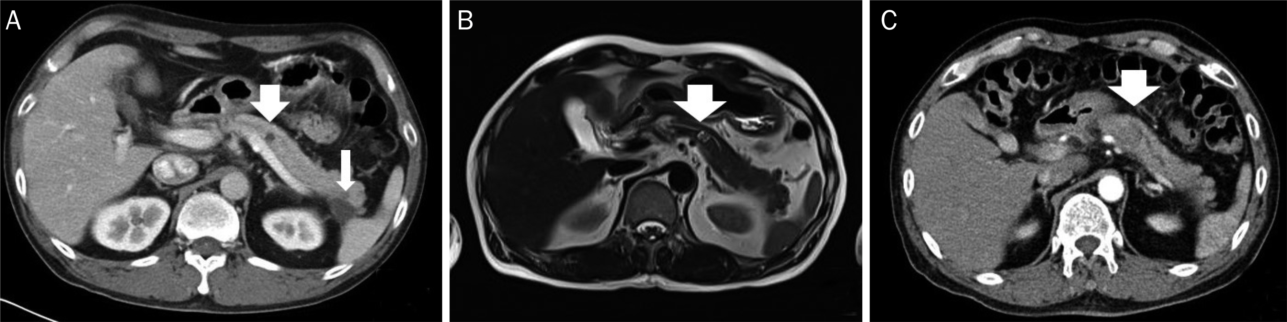
Fig. 1. (A) Two cystic masses in the pancreas body (0.8 cm, thick arrow) and tail (2.0 cm, thin arrow) were seen on contrast enhanced abdominal CT 8 months prior. (B) About 1.2 cm sized cystic mass in the pancreatic body, connected with main P-duct was seen on MRI (T1) after 3 months. The cystic mass at the pancreas tail was resorbed. (C) Hemorrhage in the 2.0 cm cystic mass of the pancreatic body was seen on the contrastenhanced abdominal CT. CT, computed tomography; MRI, magnetic resonance imaging.

Fig. 2. Blood was detected from the major papilla by esophagogastroduodenoscopy.
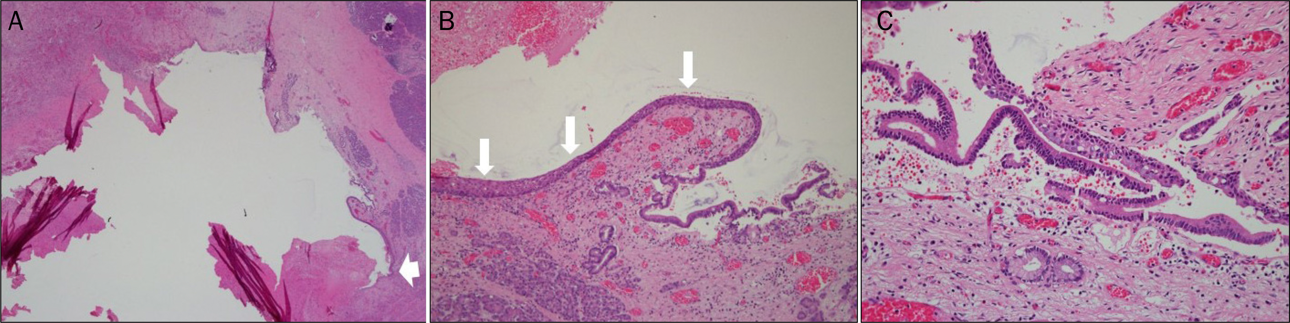
Fig. 3. (A) No papillary epithelium is seen. The normal epithelium layer is destroyed (thick arrow) (H&E, ×50). (B) Partial squamous metaplasia is seen (arrows) (H&E, ×100). (C) Mucinous epithelium without ovarian-type stroma is seen (H&E, ×200).
Reference
-
References
1. Krasinskas AM, Oakley GJ, Bagci P, et al. "Simple mucinous cyst" of the pancreas: a clinicopathologic analysis of 39 examples of a diagnostically challenging entity distinct from intraductal abdominal mucinous neoplasms and mucinous cystic neoplasms. Am J Surg Pathol. 2017; 41:121–127.2. Suter M, Doenz F, Chapuis G, Gillet M, Sandblom P. Haemorrhage into the pancreatic duct (hemosuccus pancreaticus): abdominal and management. Eur J Surg. 1995; 161:887–892.3. Risti B, Marincek B, Jost R, Decurtins M, Ammann R. abdominal pancreaticus as a source of obscure upper gastrointestinal bleeding: three cases and literature review. Am J Gastroenterol. 1995; 90:1878–1880.4. Kurland J, Matthews T, Hoff E, Gentry A, Cash B. Hemosuccus pancreaticus caused by metastatic renal cell carcinoma. Gastrointest Endosc. 2007; 66:1241–1242.
Article5. Shinzeki M, Hori Y, Fujino Y, et al. Mucinous cystic neoplasm of the pancreas presenting with hemosuccus pancreaticus: report of a case. Surg Today. 2010; 40:470–473.
Article6. Inoue H, Katurahara M, Hamada Y, et al. Hemosuccus abdominalus caused by in situ carcinoma of the pancreas. Endoscopy. 2012; 44(Suppl 2 UCTN):E336–E337.7. Kosmahl M, Egawa N, Schröder S, Carneiro F, Lüttges J, Klöppel G. Mucinous nonneoplastic cyst of the pancreas: a novel abdominal cystic change? Mod Pathol. 2002; 15:154–158.8. Vege SS, Ziring B, Jain R, Moayyedi P; Clinical Guidelines Committee; American Gastroenterology Association. American gastroenterological association institute guideline on the abdominal and management of asymptomatic neoplastic pancreatic cysts. Gastroenterology. 2015; 148:819–822. quize 12–13.
- Full Text Links
-
- Actions
-
Cited
- CITED
-
- Close
- Share
-
- Similar articles
-
- Hemosuccus Pancreaticus due to Intraductal Pseudoaneurysm
- A Case of a Choledochal Cyst with a Mucinous Cystadenoma of the Pancreas
- Two Difficultly Diagnosed Cases with Pseudoaneurysm in Chronic Pancreatitis Pseudoaneurysms Identified Inadvertently during Percutaneous Drainage and Hemosuccus Pancreaticus
- Hemosuccus Pancreaticus (Hemoductal Pancreatitis, Gastrointestinal Hemorrhage Due to Rupture of a Splenic Artery Aneurysm into the Pancreatic Duct)
- Recurrent Upper Gastrointestinal Hemorrhage due to Hemosuccus Pancreaticus from True Splenic Artery Aneurysm
