A Case of 2-Month-Old Infant with Lobular Capillary Hemangioma
- Affiliations
-
- 1Department of Otorhinolaryngology, Seoul National University College of Medicine, Seoul, Korea. handh@snu.ac.kr
- KMID: 2398832
- DOI: http://doi.org/10.18787/jr.2017.24.2.127
Abstract
- Lobular capillary hemangioma (LCH) in the nasal cavity, previously known as pyogenic granuloma, is an extremely rare benign vascular tumor in infants. LCH is a rapidly growing lesion that has a bleeding tendency due to its excessive vascularity. The authors experienced a case of LCH of the nasal cavity in a 2-month-old infant that was totally resected via the endoscopic approach after preoperative embolization. Therefore, we report this case with a brief review of the literature.
MeSH Terms
Figure
-

Fig. 1 Preoperative and postoperative photographs of the patient's face and left nasal cavity. No swelling or mass in the face (A), but left nasal cavity mass was detected (B). Protruding mass was observed after 1 month (C). The patient's postoperative 2-month follow-up photograph (D) and endoscopic image (E) shows no evidence of tumor recurrence.

Fig. 2 MRI reveals that 4×2.4 cm sized T1 low signal (A), T2 high signal (B), enhancing T1 axial (C) and coronal (D) mass in left nasal cavity.
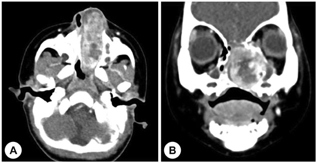
Fig. 3 Preoperative CT [Axial (A) and coronal (B) image] shows increased size (5.2×3 cm) of well-defined heterogeneous enhancing mass in the left nasal cavity.
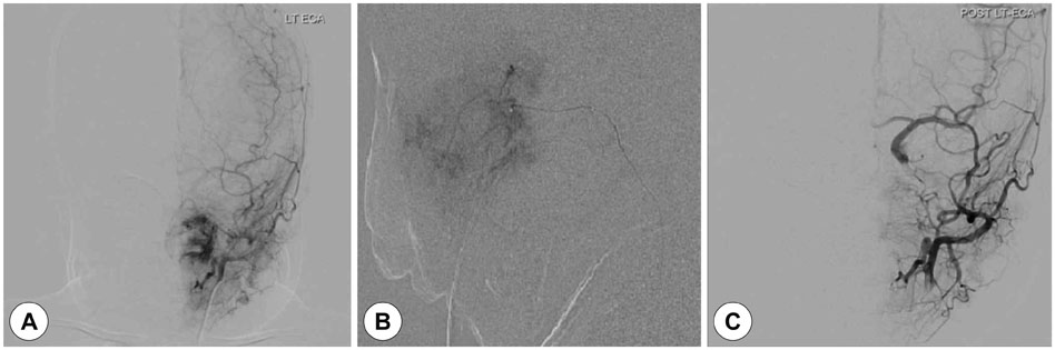
Fig. 4 Preoperative transfemoral cerebral angiogram (TFCA) and tumor embolization. The picture (A) is pre-procedure image which shows hypervascular lesion on the left nasal cavity. The picture (B) is the photo under the procedure which contrast media and embolization material is mixed and injected at the same time. The picture (C) is post-procedure image and shows devascularization of the tumor.

Fig. 5 Intraoperative endoscopic photographs. During the operation, the mass is well encapsulated and internal decompression performed using microdebrider (A). Tumor and origin site are totally resected (B). After the operation, grossly no tumor is observed (C).
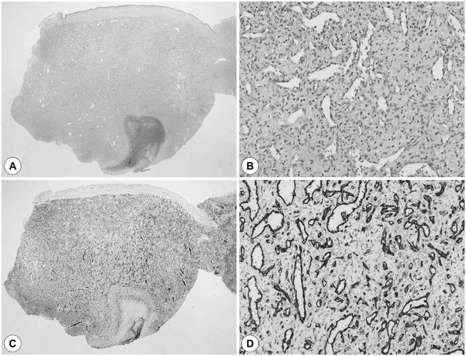
Fig. 6 Histopathologic findings of the excised mass. Thin epidermis at top with variable ulceration and lobular pattern (A) of vascular proliferation with inflammation and edema resembling granulation tissue (B) are noted. Immunohistochemical staining for CD31 reveals positivity in the endothelium of the vessels (C), (D) [original magnification ×12.5 (A), (C) and ×400 (B and D)].
Reference
-
1. Mills SE, Cooper PH, Fechner RE. Lobular capillary hemangioma: the underlying lesion of pyogenic granuloma. A study of 73 cases from the oral and nasal mucous membranes. Am J Surg Pathol. 1980; 4(5):470–479.2. el-Sayed Y, al-Serhani A. Lobular capillary haemangioma (pyogenic granuloma) of the nose. J Laryngol Otol. 1997; 111(10):941–945.
Article3. Lee JY, Koh ES. A case of cavernous hemangioma originating from the posterior end of inferior turbinate misdiagnosed as the nasopharyngeal anagiofibroma. J Rhinol. 2007; 14(1):60–64.4. Kim NY, Choi SM, Kim CH. Lobular capillary hemangioma (Pyogenic Granuloma). Korean J Otolaryngol-Head Neck Surg. 1994; 37(6):1293–1297.5. van Vugt LJ, van der Vleuten CJM, Flucke U, Blokx WAM. The utility of GLUT1 as a diagnostic marker in cutaneous vascular anomalies: A review of literature and recommendations for daily practice. Pathol Res Pract. 2017; 213(6):591–597.
Article6. Park SK, Cho HW, Jang SH, Park CK. Clinical study of lobular capillary hemangioma in nasal cavity. Korean J Otolaryngol-Head Neck Surg. 2000; 43(4):402–405.7. Lance E, Schatz C, Nach R, Thomas P. Pyogenic granuloma gravidarum of the nasal fossa: CT features. J Comput Assist Tomogr. 1992; 16(4):663–664.8. Kim KJ, Kim TY, Kim YH, Jang TY. A case of giant nasal lobular capillary hemangioma during pregnancy. J Rhinol. 2011; 18(2):142–145.9. Leyden JJ, Master GH. Oral cavity pyogenic granuloma. Arch Dermatol. 1973; 108(2):226–228.
Article10. Park MJ, Ko B, Han EM, Sohn JH. A case of multiple lobular capillary hemangiomas of the nasal septum. Korean J Otorhinolaryngol-Head Neck Surg. 2015; 58(6):421–424.
Article11. Katori H, Tsukuda M. Lobular capillary hemangioma of the nasal cavity in child. Auris Nasus Larynx. 2005; 32(2):185–188.
Article12. Kim JH, Park SW, Kim SC, Lim MK, Jang TY, Kim YJ, et al. Computed tomography and magnetic resonance imaging findings of nasal cavity hemangiomas according to histological type. Korean J Radiol. 2015; 16(3):566–574.
Article13. Chang DS, Choi MS, Lee HY, Cho CS, Park SG, Park NS, et al. Clinical study of the intranasal hemangioma. Korean J Otorhinolaryngol-Head Neck Surg. 2015; 58(5):324–329.
Article
- Full Text Links
-
- Actions
-
Cited
- CITED
-
- Close
- Share
-
- Similar articles
-
- A Rare Case of Synchronous Pleomorphic Adenoma and Lobular Capillary Hemangioma of Nasal Septum
- A Case of Concurrent Osteoma and Lobular Capillary Hemangioma of the Middle Turbinate
- A Case of Lobular Capillary Hemangioma at the False Vocal Cord With Intermittent Stridor
- A Rare Case of Synchronous Lobular Capillary Hemangioma and Non-Lobular Capillary Hemangioma Arising From the Both Arytenoid Mimicking Laryngeal Granuloma
- Subcutaneous Pyogenic Granuloma (Lobular Capillary Hemangioma) on the Nose Mistaken for Sebaceous Gland Hyperplasia
