A Case of Fungal Ball Caused by Retained Glass Foreign Body in Maxillary Sinus for 30 Years
- Affiliations
-
- 1Department of Otorhinolaryngology-Head and Neck Surgery, School of Medicine, Kyungpook National University, Daegu, Korea. profsookim@gmail.com
- KMID: 2398831
- DOI: http://doi.org/10.18787/jr.2017.24.2.123
Abstract
- The fungal ball is the most frequent non-invasive form of fungal sinusitis. Some authors have reported opportunistic fungus infections caused by retained foreign bodies in the maxillary sinus. Whereas previously reported foreign bodies were almost always metal materials, we report the case of a fungus ball caused by retained pieces of glass for 30 years. The patient complained of unilateral nasal obstruction. Computed tomography revealed fungal sinusitis and foreign bodies in the left maxillary sinus. The fungus ball and foreign bodies were removed via an endoscopic sinus surgery. After surgical removal, the patient became free from nasal symptoms.
Keyword
Figure
-
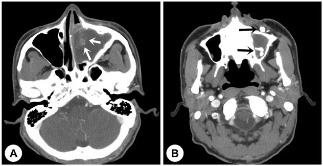
Fig. 1 Preoperative axial computed tomography scan. A: Calcific density (white arrow) is shown in the left maxillary sinus. B: Foreign bodies (black arrow) are shown in the floor of the left maxillary sinus and anterior to the left maxilla.
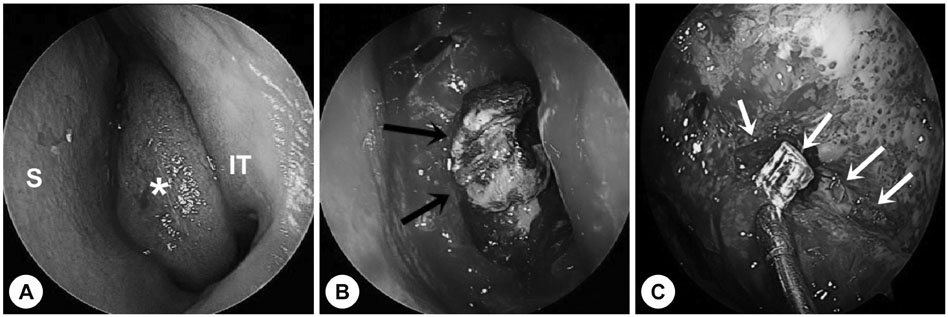
Fig. 2 Intraoperative endoscopic findings. A : Uncinate process and medial wall of left maxillary sinus (*) extended to septum (S). B and C : Fungal ball like materials (black arrow) and several pieces of glass (white arrow) are shown in the left maxillary sinus. IT, inferior turbinate.
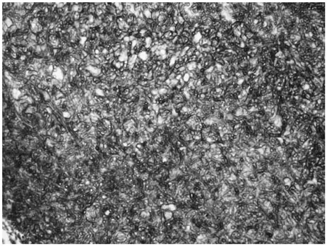
Fig. 3 The numerous septated fungal hyphaes are observed in GMS stain (×40). The fungal hyphaes show septate filaments, regular thick, branching at acute angle. These morphologic findings are consistent with Aspergillus. GMS: Gomori's methenamine silver.
Reference
-
1. Burnham R, Bridle C. Aspergillosis of the maxillary sinus secondary to a foreign body (amalgam) in the maxillary antrum. Br J Oral Maxillofac Surg. 2009; 47:313–315.
Article2. Kobayashi A. Asymptomatic aspergillosis of the maxillary sinus associated with foreign body of endodontic origin. Report of a case. Int J Oral Maxillofac Surg. 1995; 24:243–244.
Article3. Chung YJ. Secondary Fungus Ball Caused by a Retained Foreign Body in Maxillary Sinus. Korean J Otorhinolaryngol-Head Neck Surg. 2014; 57:132–135.
Article4. Lee JH, Kim JM, Jeong HM, Lee SH. A Case of Fungal Ball Accompanied with a Microplate as Metallic Foreign Body in Maxillary Sinus. Korean J Otorhinolaryngol-Head Neck Surg. 2013; 56:735–737.
Article5. Lee DH, Lim SC. Maxillary fungus ball caused by retained foreign bodies for 25 years. J Craniofac Surg. 2012; 23:e176–e177.
Article6. Kim DH, Park TJ, Kwon J, Kim JG. A Case of Foreign Body Incidentally Found at the Maxilla and Maxillary Sinus. Korean J Otorhinolaryngol-Head Neck Surg. 2012; 55:787–790.
Article7. Guidera AK, Dixon PM, Stegehuis HR. Glass in the frontal sinus: 28-year delayed presentation. Ear Nose Throat J. 2013; 92:E10.8. Kim JH, Kim SH, Kim JH, Kim KS. A Case of Metallic Foreign Body Retained in the Naso-Maxillo-Ethmoido-Orbital Complex. Korean J Otolaryngol-Head Neck Surg. 2004; 47:177–181.9. Legent F, Billet J, Beauvillain C, Bonnet J, Miegeville M. The role of dental canal fillings in the development of Aspergillus sinusitis. A report of 85 cases. Arch Otorhinolaryngol. 1989; 246:318–320.
Article10. Stammberger H. Endoscopic surgery for mycotic and chronic recurring sinusitis. Ann Otol Rhinol Laryngol Suppl. 1985; 119:1–11.
Article11. Jeong JH, Jeon SY, Ahn SK, Cho HG. A Case of Frontal Mucocele with Paranasal Sinus Glass Foreign Body in Head Trauma Patient. J Rhinol. 2005; 12:112–115.12. Onerci M, Ogretmenoglu O, Yilmaz T. Glass in the frontal sinus: report of three cases. J Laryngol Otol. 1997; 111:156–158.
Article13. Yeo CK, Ahn BH, Kim JS, Kim YD, Shin SH, Ye MK. Fungal Ball in Sinus: Multi-Center Study in Daegu. J Rhinol. 2005; 12:105–107.
- Full Text Links
-
- Actions
-
Cited
- CITED
-
- Close
- Share
-
- Similar articles
-
- Secondary Fungus Ball Caused by a Retained Foreign Body in Maxillary Sinus
- A Case of Fungal Ball Accompanied with a Microplate as Metallic Foreign Body in Maxillary Sinus
- A Case of Actinomycosis of the Maxillary Sinus Accompanied with Fungal Ball
- Long-Standing Blades of Scissors in Paranasal Sinus Over 40 Years
- A Case of Foreign Body Incidentally Found at the Maxilla and Maxillary Sinus
