J Adv Prosthodont.
2017 Dec;9(6):439-446. 10.4047/jap.2017.9.6.439.
Influence of different surface treatments on bond strength of novel CAD/CAM restorative materials to resin cement
- Affiliations
-
- 1Department of Prosthodontics, School of Dentistry, Ordu University, Turkey. aycatulga@odu.edu.tr
- KMID: 2398048
- DOI: http://doi.org/10.4047/jap.2017.9.6.439
Abstract
- PURPOSE
To evaluate the effects of different surface treatments on the bond strength of novel CAD/CAM restorative materials to resin cement by four point bending test.
MATERIALS AND METHODS
The CAD/CAM materials under investigation were e.max CAD, Mark II, Lava Ultimate, and Enamic. A total of 400 bar specimens (4×1.2×12 mm) (n=10) milled from the CAD/CAM blocks underwent various pretreatments (no pretreatment (C), hydrofluoric acid (A), hydrofluoric acid + universal adhesive (Scotchbond) (AS), sandblasting (Sb), and sandblasting + universal adhesive (SbS)). The bars were luted end-to-end on the prepared surfaces with a dual curing adhesive resin cement (Variolink N, Ivoclar Vivadent) on the custom-made stainless steel mold. Ten test specimens for each treatment and material combination were performed with four point bending test method. Data were analyzed using ANOVA and Tukey's test.
RESULTS
The surface treatment and type of CAD/CAM restorative material showed a significant effect on the four point bending strength (FPBS) (P < .001). For LDC, AS surface treatment showed the highest FPBS results (100.31 ± 10.7 MPa) and the lowest values were obtained in RNC (23.63 ± 9.0 MPa) for control group. SEM analyses showed that the surface topography of CAD/CAM restorative materials was modified after treatments.
CONCLUSION
The surface treatment of sandblasting or HF acid etching in combination with a universal adhesive containing MDP can be suggested for the adhesive cementation of the novel CAD/CAM restorative materials.
Keyword
MeSH Terms
Figure
-
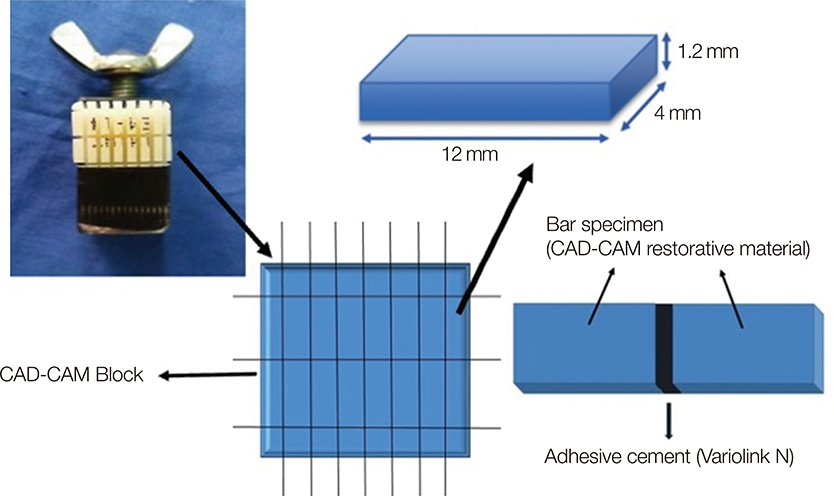
Fig. 1 Schematic drawing illustrating the study set-up and specimen preparation procedure.
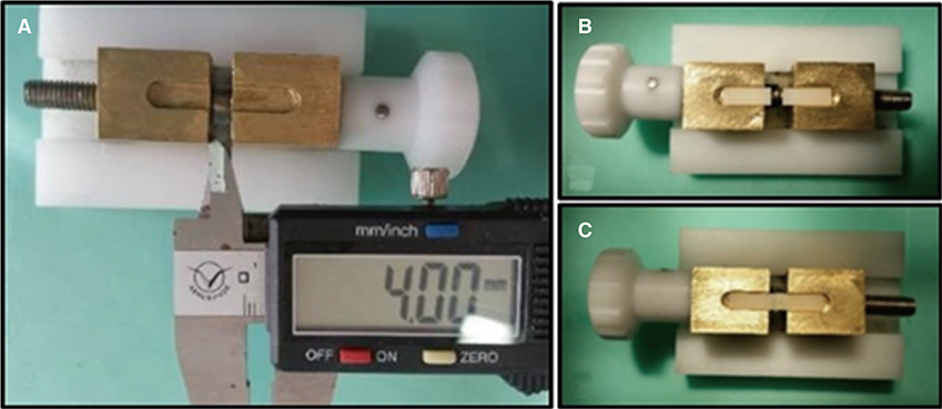
Fig. 2 (A) A custom made stainless steel mold was used for cementation. After screwing down the instrument for cementation, the distance of separated two fragments was 4 mm; (B) Setting the specimens end to end to the mold; (C) Cementation of a pair of specimens.
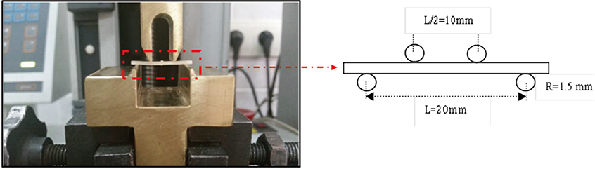
Fig. 3 The custom made device for four point bending test and experimental setup of the test.
Cited by 1 articles
-
Wear of contemporary dental composite resin restorations: a literature review
Dimitrios Dionysopoulos, Olga Gerasimidou
Restor Dent Endod. 2021;46(2):e18. doi: 10.5395/rde.2021.46.e18.
Reference
-
1. Li RW, Chow TW, Matinlinna JP. Ceramic dental biomaterials and CAD/CAM technology: state of the art. J Prosthodont Res. 2014; 58:208–216.2. Mainjot AK, Dupont NM, Oudkerk JC, Dewael TY, Sadoun MJ. From Artisanal to CAD-CAM Blocks: State of the Art of Indirect Composites. J Dent Res. 2016; 95:487–495.3. Miyazaki T, Hotta Y. CAD/CAM systems available for the fabrication of crown and bridge restorations. Aust Dent J. 2011; 56:97–106.4. Della Bona A, Nogueira AD, Pecho OE. Optical properties of CAD-CAM ceramic systems. J Dent. 2014; 42:1202–1209.5. Ruse ND, Sadoun MJ. Resin-composite blocks for dental CAD/CAM applications. J Dent Res. 2014; 93:1232–1234.6. Bonfante EA, Suzuki M, Lorenzoni FC, Sena LA, Hirata R, Bonfante G, Coelho PG. Probability of survival of implant-supported metal ceramic and CAD/CAM resin nanoceramic crowns. Dent Mater. 2015; 31:e168–e177.7. Schepke U, Meijer HJ, Vermeulen KM, Raghoebar GM, Cune MS. Clinical bonding of resin nano ceramic restorations to zirconia abutments: A case series within a randomized clinical trial. Clin Implant Dent Relat Res. 2016; 18:984–992.8. Coldea A, Swain MV, Thiel N. In-vitro strength degradation of dental ceramics and novel PICN material by sharp indentation. J Mech Behav Biomed Mater. 2013; 26:34–42.9. Coldea A, Swain MV, Thiel N. Mechanical properties of polymer-infiltrated-ceramic-network materials. Dent Mater. 2013; 29:419–426.10. Della Bona A, Corazza PH, Zhang Y. Characterization of a polymer-infiltrated ceramic-network material. Dent Mater. 2014; 30:564–569.11. Frankenberger R, Taschner M, Garcia-Godoy F, Petschelt A, Krämer N. Leucite-reinforced glass ceramic inlays and onlays after 12 years. J Adhes Dent. 2008; 10:393–398.12. Reiss B. Clinical results of Cerec inlays in a dental practice over a period of 18 years. Int J Comput Dent. 2006; 9:11–22.13. Roggendorf MJ, Kunzi B, Ebert J, Roggendorf HC, Frankenberger R, Reich SM. Seven-year clinical performance of CEREC-2 all-ceramic CAD/CAM restorations placed within deeply destroyed teeth. Clin Oral Investig. 2012; 16:1413–1424.14. Sjögren G, Molin M, van Dijken JW. A 10-year prospective evaluation of CAD/CAM-manufactured (Cerec) ceramic inlays cemented with a chemically cured or dual-cured resin composite. Int J Prosthodont. 2004; 17:241–246.15. Thordrup M, Isidor F, Hörsted-Bindslev P. A prospective clinical study of indirect and direct composite and ceramic inlays: ten-year results. Quintessence Int. 2006; 37:139–144.16. Frankenberger R, Hartmann VE, Krech M, Krämer N, Reich S, Braun A, Roggendorf M. Adhesive luting of new CAD/CAM materials. Int J Comput Dent. 2015; 18:9–20.17. El Zohairy AA, De Gee AJ, Mohsen MM, Feilzer AJ. Microtensile bond strength testing of luting cements to prefabricated CAD/CAM ceramic and composite blocks. Dent Mater. 2003; 19:575–583.18. Frankenberger R, Lohbauer U, Taschner M, Petschelt A, Nikolaenko SA. Adhesive luting revisited: influence of adhesive, temporary cement, cavity cleaning, and curing mode on internal dentin bond strength. J Adhes Dent. 2007; 9:269–273.19. Dos Santos VH, Griza S, de Moraes RR, Faria-E-Silva AL. Bond strength of self-adhesive resin cements to composite submitted to different surface pretreatments. Restor Dent Endod. 2014; 39:12–16.20. Elsaka SE. Effect of surface treatments on the bonding strength of self-adhesive resin cements to zirconia ceramics. Quintessence Int. 2013; 44:407.21. Li R. Development of a ceramic primer with higher bond durability for resin cement. J Oral Rehabil. 2010; 37:560–568.22. Rosa WL, Piva E, Silva AF. Bond strength of universal adhesives: A systematic review and meta-analysis. J Dent. 2015; 43:765–776.23. Elsaka SE. Bond strength of novel CAD/CAM restorative materials to self-adhesive resin cement: the effect of surface treatments. J Adhes Dent. 2014; 16:531–540.24. Şanlı S, Çömlekoğlu MD, Çömlekoğlu E, Sonugelen M, Pamir T, Darvell BW. Influence of surface treatment on the resinbonding of zirconia. Dent Mater. 2015; 31:657–668.25. Touaiher I, Saâdaoui M, Chevalier J, Reveron H. Effect of loading configuration on strength values in a highly transformable zirconia-based composite. Dent Mater. 2016; 32:e211–e219.26. Yassini E, Mirzaei M, Alimi A, Rahaeifard M. Investigation of the fatigue behavior of adhesive bonding of the lithium disilicate glass ceramic with three resin cements using rotating fatigue method. J Mech Behav Biomed Mater. 2016; 61:62–69.27. Peumans M, Valjakova EB, De Munck J, Mishevska CB, Van Meerbeek B. Bonding Effectiveness of Luting Composites to Different CAD/CAM Materials. J Adhes Dent. 2016; 18:289–302.28. Queiroz JR, Souza RO, Nogueira Junior L Jr, Ozcan M, Bottino MA. Influence of acid-etching and ceramic primers on the repair of a glass ceramic. Gen Dent. 2012; 60:e79–e85.29. Schwenter J, Schmidli F, Weiger R, Fischer J. Adhesive bonding to polymer infiltrated ceramic. Dent Mater J. 2016; 35:796–802.30. Della Bona A, van Noort R. Shear vs. tensile bond strength of resin composite bonded to ceramic. J Dent Res. 1995; 74:1591–1596.31. Valandro LF, Ozcan M, Amaral R, Vanderlei A, Bottino MA. Effect of testing methods on the bond strength of resin to zirconia-alumina ceramic: microtensile versus shear test. Dent Mater J. 2008; 27:849–855.32. Ban S, Anusavice KJ. Influence of test method on failure stress of brittle dental materials. J Dent Res. 1990; 69:1791–1799.33. Fischer J, Grohmann P, Stawarczyk B. Effect of zirconia surface treatments on the shear strength of zirconia/veneering ceramic composites. Dent Mater J. 2008; 27:448–454.34. Lima JM, Anami LC, Rippe MP, de Melo RM, Bottino MA, Valera MC, Araújo MA. Surface agents' influence on the flexural strength of bilaminated ceramics. Braz Oral Res. 2013; 27:311–317.35. Aboushelib MN, Sleem D. Microtensile bond strength of lithium disilicate ceramics to resin adhesives. J Adhes Dent. 2014; 16:547–552.36. Cekic-Nagas I, Ergun G, Egilmez F, Vallittu PK, Lassila LV. Micro-shear bond strength of different resin cements to ceramic/glass-polymer CAD-CAM block materials. J Prosthodont Res. 2016; 60:265–273.37. Reich SM, Wichmann M, Frankenberger R, Zajc D. Effect of surface treatment on the shear bond strength of three resin cements to a machinable feldspatic ceramic. J Biomed Mater Res B Appl Biomater. 2005; 74:740–746.38. Blatz MB, Phark JH, Ozer F, Mante FK, Saleh N, Bergler M, Sadan A. In vitro comparative bond strength of contemporary self-adhesive resin cements to zirconium oxide ceramic with and without air-particle abrasion. Clin Oral Investig. 2010; 14:187–192.39. Souza RO, Castilho AA, Fernandes VV, Bottino MA, Valandro LF. Durability of microtensile bond to nonetched and etched feldspar ceramic: self-adhesive resin cements vs conventional resin. J Adhes Dent. 2011; 13:155–162.40. Brentel AS, Ozcan M, Valandro LF, Alarça LG, Amaral R, Bottino MA. Microtensile bond strength of a resin cement to feldpathic ceramic after different etching and silanization regimens in dry and aged conditions. Dent Mater. 2007; 23:1323–1331.41. Toledano M, Osorio R, Osorio E, Aguilera FS, Yamauti M, Pashley DH, Tay F. Durability of resin-dentin bonds: effects of direct/indirect exposure and storage media. Dent Mater. 2007; 23:885–892.42. Pollington S, Fabianelli A, van Noort R. Microtensile bond strength of a resin cement to a novel fluorcanasite glass-ceramic following different surface treatments. Dent Mater. 2010; 26:864–872.
- Full Text Links
-
- Actions
-
Cited
- CITED
-
- Close
- Share
-
- Similar articles
-
- The effects of different surface treatments on the shear bond strengths of two dual-cure resin cements to CAD/CAM restorative materials
- Shear bond strength between CAD/CAM denture base resin and denture artificial teeth when bonded with resin cement
- Influence of surface treatments and repair materials on the shear bond strength of CAD/CAM provisional restorations
- Repair bond strength of resin composite to three aged CAD/CAM blocks using different repair systems
- Shear bond strength of the three different kinds of resin cement on CAD/CAM ceramic inlay
