J Endocr Surg.
2017 Dec;17(4):149-152. 10.16956/jes.2017.17.4.149.
Retained Drainage Tube Fragment after Thyroidectomy Presenting as a Palpable Neck Mass: a Case Report
- Affiliations
-
- 1Department of Surgery, Chonnam National University Medical School, Gwangju, Korea. mhpark@chonnam.ac.kr
- 2Department of Radiology, Chonnam National University Medical School, Gwangju, Korea.
- 3Department of Pathology, Chonnam National University Medical School, Gwangju, Korea.
- KMID: 2397991
- DOI: http://doi.org/10.16956/jes.2017.17.4.149
Abstract
- A retained drainage tube fragment after surgery is a rare complication with potentials for medical dispute. We report the case of a retained drainage tube fragment in the anterior neck following conventional thyroid surgery. To the best of our knowledge, this is the first report of its kind. A 61-year-old female patient who was found to have a retained Penrose drainage tube after thyroid surgery for a papillary thyroid cancer. The retained tube was detected 18 months after surgery presenting as a palpable neck mass. Neck ultrasonography revealed a hypoechoic lesion with tubular-shaped parallel hyperechoic lines, suggesting a retained drain tube. The lateral soft tissue neck radiograph demonstrated a radio-opaque drain tube in the anterior neck. The fragment of a Penrose drainage tube was neglected first, and then detected as a palpable neck mass 18 months after surgery. Removal of the drainage tube fragment was performed under local anesthesia. After the surgery, the operation site healed without any complications. Although drainage procedure is regarded to be easy, there can be associated complications including retained drain, bleeding, infection, or cosmetic problem. So, clinicians should be aware of rare complications associated with Penrose drainage tubes after thyroidectomy and perform this procedure carefully and gently to avoid them.
Keyword
MeSH Terms
Figure
-

Fig. 1 The longitudinal neck ultrasonography shows a hypoechoic lesion with internal echogenic parallel lines (arrows), suggesting a retained drainage tube.
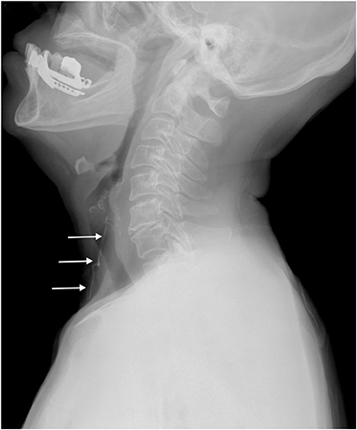
Fig. 2 The lateral soft tissue neck radiograph displays a Penrose drain fragment (arrows) in the anterior neck.
Reference
-
1. Samraj K, Gurusamy KS. Wound drains following thyroid surgery. Cochrane Database Syst Rev. 2007; CD006099.
Article2. Tabaqchali MA, Hanson JM, Proud G. Drains for thyroidectomy/parathyroidectomy: fact or fiction? Ann R Coll Surg Engl. 1999; 81:302–305.3. McCullough BS, Witherington JD. Retained penrose drain following cholecystectomy. JAMA. 1973; 225:415–416.
Article4. Karçaaltincaba M, Demirkazik FB, Imamoğlu T, Firat A, Ariyürek M. Breast abscess mimicking malignant mass due to retained penrose drain: diagnosis by mammography and MRI. Clin Imaging. 2004; 28:278–279.5. Takeyama S, Aoki Y, Kamimura N, Suzuki M, Tanaka K. Retrieval of intraperitoneal penrose drain under transvaginal endoscopic guidance. Gynecol Oncol. 2006; 102:391–393.
Article6. Liu KS, Huang KC, Wong CH. A neglected retained penrose drain mimicking an amputation stump neuroma. J Trauma. 2007; 62:1051–1052.
Article7. Leonovicz PF, Uehling DT. Removal of retained penrose drain under fluoroscopic guidance. Urology. 1999; 53:1221.
Article8. Chung YE, Kim EK, Kim MJ, Yun M, Hong SW. Suture granuloma mimicking recurrent thyroid carcinoma on ultrasonography. Yonsei Med J. 2006; 47:748–751.
Article
- Full Text Links
-
- Actions
-
Cited
- CITED
-
- Close
- Share
-
- Similar articles
-
- Apparent Sparganosis Presenting as a Palpable Neck Mass: A Case Report and Review of Literature
- Airway Obstruction by Swelling of Inner Wall of Armored Tube and Tracheal Laceration during Endoscopic Thyroidectomy: A case report
- Intraoperative Neuromonitoring with Adhesive Skin Electrodes for a Patient with Papillary Thyroid Carcinoma with Lateral Neck Metastasis: a Case Report
- Bilateral Chylothorax Following Modified Radical Neck Dissection in Thyroid Cancer: A Case Report
- Dacryocystorhinostomy with Silicone Tube used in the Lacrimal Drainage System
