Anterior Segment Changes after Laser Iridotomy for the Treatment and Prevention of Angle-closure Glaucoma
- Affiliations
-
- 1Department of Ophthalmology, Cheonan Hospital, Soonchunhyang University College of Medicine, Cheonan, Korea. inmydream@schmc.ac.kr
- KMID: 2397854
- DOI: http://doi.org/10.3341/jkos.2017.58.12.1396
Abstract
- PURPOSE
To evaluate the changes and characteristics of the anterior segment of the eye after laser peripheral iridotomy (LPI) conducted on patients with acute angle closure crisis (AACC) for both therapeutic purposes and prophylactic purposes in the fellow eye.
METHODS
Anterior segments were examined by topography, laser interferometry, anterior segment optical coherence tomography, anterior chamber depth (ACD), anterior chamber volume (ACV), anterior chamber angle (ACA), angle opening distance (AOD), central corneal thickness (CCT), and axial length as compared to prior procedures in 20 eyes with glaucoma (treatment group) and 20 contralateral eyes (prophylactic group) in 20 patients diagnosed with AACC.
RESULTS
Before laser treatment, there were no significant differences in pre-LPI ACV, ACA, AOD and axial length, although differences in the CCT and ACD existed between the groups. Compared to prior laser treatment at 1 and 3 months after laser treatment, the ACV, ACA, AOD 500, and AOD 750 increased in both groups. When both groups were compared 1 month after their laser treatments, the AOD 750 was increased in the treatment group. There were no significant differences between two groups 3 months post LPI.
CONCLUSIONS
Other than changes in the ACD and CCT, no significant differences were observed in the anterior segment characteristics in AACC affected and contralateral eyes prior to LPI. After LPI, the treatment group showed greater changes in their anterior segments; however, the open angle was maintained at 1 month post treatment.
Keyword
MeSH Terms
Figure
-
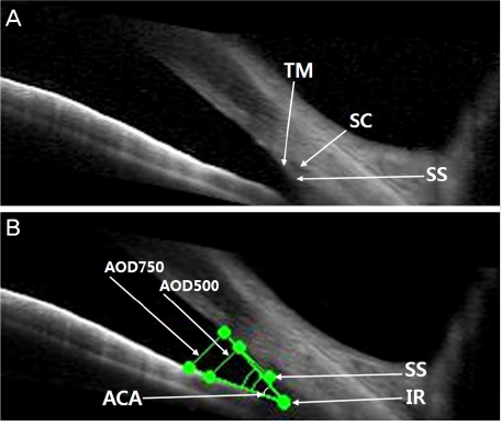
Figure 1 Anterior chamber image of anterior segment optical coherence tomography. (A, B) anterior chamber angle (ACA) means apex in the iris recess (IR) and the arms of the angle passing through a point on the trabecular meshwork (TM) from the scleral spur and the point on the iris perpendicularly opposite, angle opening distance (AOD) 500, 750 means distance between the point which was 500 µm (750 µm) from anterior to the scleral spur (SS) and the iris (perpendicularly opposite point). SC = Schlemm's canal.

Figure 2 Changes in anterior segment parameters over time in treatment group and prophylactic group. (A) Angle opening distance 750 µm (AOD 750), (B) anterior chamber angle (ACA) (Confidence interval = 95%). Before laser treatment, there were no significant differences in pre- laser peripheral iridotomy (LPI) AOD 750, ACA between two groups. Compared with prior laser treatment, 1 month and 3 months after laser treatment, the AOD 750 and ACA was increased in both groups. When two groups were compared 1 month after the laser treatment, the change was greater in the treatment group. And no significant differences between two groups after 3 months of LPI.
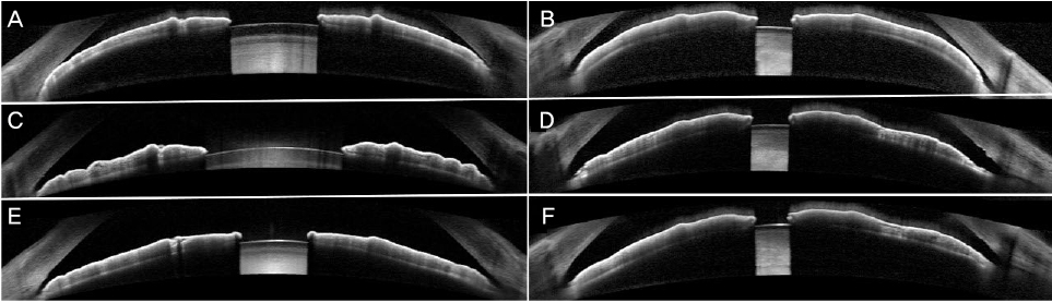
Figure 3 Laser peripheral iridotomy (LPI) in patient with acute angle closure crisis (AACC). Pre-LPI anterior segment optical coherence tomography of a 70-year-old female with AACC on the right eye, showing closed angles (A) and the left eye (B). (C, D) Horizontal section scan showing increased anterior chamber angle and angle opening distance after 1 month from LPI. (E, F) Horizontal section scan after 3 months from LPI.
Reference
-
1. Thylefors B, Négrel AD. The global impact of glaucoma. Bull World Health Organ. 1994; 72:323–326.2. Dandona L, Dandona R, Mandal P, et al. Angle-closure glaucoma in an urban population in southern India. The Andhra Pradesh eye disease study. Ophthalmology. 2000; 107:1710–1716.3. Foster PJ, Johnson GJ. Glaucoma in China: how big is the problem? Br J Ophthalmol. 2001; 85:1277–1282.4. Chew PT, Aung T. Primary angle-closure glaucoma in Asia. J Glaucoma. 2001; 10:5 Suppl 1. S7–S8.5. Alsbirk PH. Anterior chamber depth, genes and environment. A population study among long-term Greenland Eskimo immigrants in Copenhagen. Acta Ophthalmol (Copenh). 1982; 60:223–224.6. Sihota R, Lakshmaiah NC, Agarwal HC, et al. Ocular parameters in the subgroups of angle closure glaucoma. Clin Exp Ophthalmol. 2000; 28:253–258.7. Congdon NG, Youlin Q, Quigley H, et al. Biometry in primary angle-closure glaucoma among Chinese, white, and black populations. Ophthalmology. 1997; 104:1489–1495.8. Friedman DS, Chew PT, Gazzard G, et al. Long-term outcomes in fellow eyes after acute primary angle closure in the contralateral eye. Ophthalmology. 2006; 113:1087–1091.9. Ang LP, Aung T, Chew PT. Acute primary angle closure in an Asian population: long-term outcome of the fellow eye after prophylactic laser peripheral iridotomy. Ophthalmology. 2000; 107:2092–2096.10. Lee JR, Sung KR, Han S. Comparison of anterior segment parameters between the acute primary angle closure eye and the fellow eye. Invest Ophthalmol Vis Sci. 2014; 55:3646–3650.11. Kim DY, Sung KR, Kang SY, et al. Characteristics and reproducibility of anterior chamber angle assessment by anterior-segment optical coherence tomography. Acta Ophthalmol. 2011; 89:435–441.12. Lee Y, Sung KR, Na JH, Sun JH. Dynamic changes in anterior segment (AS) parameters in eyes with primary angle closure (PAC) and PAC glaucoma and open-angle eyes assessed using AS optical coherence tomography. Invest Ophthalmol Vis Sci. 2012; 53:693–697.13. Baek S, Sung KR, Sun JH, et al. A hierarchical cluster analysis of primary angle closure classification using anterior segment optical coherence tomography parameters. Invest Ophthalmol Vis Sci. 2013; 54:848–853.14. Huang J, Pesudovs K, Wen D, et al. Comparison of anterior segment measurements with rotating Scheimpflug photography and partial coherence reflectometry. J Cataract Refract Surg. 2011; 37:341–348.15. Grewal DS, Brar GS, Jain R, Grewal SP. Comparison of Scheimpflug imaging and spectral domain anterior segment optical coherence tomography for detection of narrow anterior chamber angles. Eye (Lond). 2011; 25:603–611.16. Kang JE, Lee HJ, Choi KR. Ultrasound biomicroscopic assessment of the changes in angle relationships after laser iridotomy in primary angle-closure glaucoma. J Korean Ophthalmol Soc. 2005; 46:800–809.17. Hong C, Joo JH, Shin KH, Song KY. Clinical study of Korean glaucomatous patients. Korean J Ophthalmol. 1987; 1:41–46.18. Thomlison A, Leighton DA. Ocular dimensions and the hereditary of angle-closure glaucoma. Br J Ophthalmol. 1973; 57:475.19. Salmon JF. Predisposing factors for chronic angle-closure glaucoma. Prog Retin Eye Res. 1999; 18:121–132.20. Ritch R, Lowe RF. Angle-closure glaucoma: mechanisms and epidemiology. In : Ritch R, Shields MB, Krupin T, editors. The Glaucomas. 2nd ed. St Louis: CV Mosby;1996. v.2:chap. 37.21. Lowe RF. Aetiology of the anatomical basis for primary angle-closure glaucoma. Biometrical comparisons between normal eyes and eyes with primary angle-closure glaucoma. Br J Ophthalmol. 1970; 54:161–169.22. Marchini G, Pagliarusco A, Toscano A, et al. Ultrasound biomicroscopic and conventional ultrasonographic study of ocular dimensions in primary angle-closure glaucoma. Ophthalmology. 1998; 105:2091–2098.23. Lowe RF. Primary creeping angle-closure glaucoma. Br J Ophthalmol. 1964; 48:544–550.24. He M, Friedman DS, GE J, et al. Laser peripheral iridotomy in eyes with narrow drainage angles: ultrasound biomicroscopy outcomes. The Liwan Eye Study. Ophthalmology. 2007; 114:1513–1519.25. Jiang Y, Chang DS, Zhu H, et al. Longitudinal changes of angle configuration in primary angle-closure sustepcts: the Zhongshan Angle-Closure Prevention Trial. Ophthalmology. 2014; 121:1699–1705.26. Lee KS, Sung KR, Shon K, et al. Longitudinal changes in anterior segment parameters after laser peripheral iridotomy assessed by anterior segment opcital coherence tomography. Invest Ophthalmol Vis Sci. 2013; 54:3166–3170.27. Loon SC, Chew PT, Oen FT, et al. Iris ischaemic changes and visual outcome after acute primary angle closure. Clin Exp Ophthalmol. 2005; 33:473–477.28. Cao JT, Wu L, Liu SY. Histopathologic and ultrastructural studies of the iris in acute angle-closure glaucoma. Zhonghua Yan Ke Za Zhi. 1994; 30:264–266.
- Full Text Links
-
- Actions
-
Cited
- CITED
-
- Close
- Share
-
- Similar articles
-
- Effect of YAG Laser Iridotomy on IOP in Chronic Angle-closure Glaucoma
- Argon Laser Iridotomy
- Clinical Results of the Q-Switched Nd-YAG Laser Iridotomy
- Effectiveness of Argon Laser Peripheral Iridoplasty in the Treatment of Severe Acute Angle-Closure Glaucoma
- Clinical Experience of Q-switched Nd:YAG Laser Iridotomy 50 cases for Angle Glosure Glaucoma
