Submandibular sialolithiasis with CT and scintigraphy: CT values and salivary gland excretion in the submandibular glands
- Affiliations
-
- 1Radiology, The Nippon Dental University Niigata Hospital, Niigata, Japan. ogura@ngt.ndu.ac.jp
- 2Department of Oral and Maxillofacial Radiology, The Nippon Dental University School of Life Dentistry at Niigata, Niigata, Japan.
- KMID: 2397836
- DOI: http://doi.org/10.5624/isd.2017.47.4.227
Abstract
- PURPOSE
Sialolithiasis is one of the most prevalent large obstructive disorders of the submandibular glands. The aim of this study was to investigate submandibular sialolithiasis with computed tomography (CT) and scintigraphy, with a particular focus on the relationship between CT values of the submandibular glands and their excretion rate.
MATERIALS AND METHODS
Fifteen patients with submandibular sialolithiasis who underwent CT and salivary gland scintigraphy were included in this study. The relationship between the CT values of submandibular glands with and without sialoliths and salivary gland excretion measured using salivary gland scintigraphy was statistically analyzed. Dynamic images were recorded on the computer at 1 frame per 20 seconds. The salivary gland excretion fraction was defined as A (before stimulation test [counts/frame]) / B(after stimulation test [counts/frame]) using time-activity curves.
RESULTS
The CT values in the submandibular glands with and without sialoliths was 9.9±44.9 Hounsfield units (HU) and 34.2±21.8 HU, respectively (P=.233). Regarding the salivary gland excretion fraction using scintigraphy, the A/B value in the submandibular glands with sialoliths (1.09±0.23) was significantly lower than in the submandibular glands without sialoliths (1.99±0.57, P=.000).
CONCLUSION
Assessments of the CT values and the salivary gland excretion fraction using scintigraphy in the submandibular glands seem to be useful tools evaluating submandibular sialolithiasis.
Keyword
MeSH Terms
Figure
-
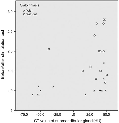
Fig. 1 Correlation between computed tomography (CT) values of submandibular glands with/without sialoliths and before/after the stimulation test. With: submandibular gland with sialoliths, without: submandibular gland without sialoliths.
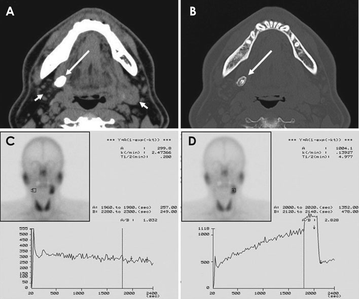
Fig. 2 A 50-year-old male with right-sided submandibular sialolithiasis. An axial soft-tissue algorithm computed tomography (CT) image (A) demonstrates a sialolith (long arrow) and submandibular glands (short arrows). The density of the right submandibular gland was less than that of the left submandibular gland. Axial bone tissue algorithm CT (B) shows a sialolith (long arrow). The A (before stimulation test [counts/frame]) / B (after stimulation test [counts/frame]) value using time-activity curves of the right submandibular gland (C) was lower than that of the left submandibular gland (D).
Reference
-
1. Kim JH, Aoki EM, Cortes AR, Abdala-Júnior R, Asaumi J, Arita ES. Comparison of the diagnostic performance of panoramic and occlusal radiographs in detecting submandibular sialoliths. Imaging Sci Dent. 2016; 46:87–92.
Article2. Lustmann J, Regev E, Melamed Y. Sialolithiasis. A survey on 245 patients and a review of the literature. Int J Oral Maxillofac Surg. 1990; 19:135–138.3. Schwarz D, Kabbasch C, Scheer M, Mikolajczak S, Beutner D, Luers JC. Comparative analysis of sialendoscopy, sonography, and CBCT in the detection of sialolithiasis. Laryngoscope. 2015; 125:1098–1101.
Article4. Gulati A, Scott J, Blythe JN, Southorn B, Brennan PA. Review of salivary papers published in the British Journal of Oral & Maxillofacial Surgery during 2009-2010. Br J Oral Maxillofac Surg. 2011; 49:627–629.5. Wu CB, Xi H, Zhou Q, Zhang LM. The diagnostic value of technetium 99m pertechnetate salivary gland scintigraphy in patients with certain salivary gland diseases. J Oral Maxillofac Surg. 2015; 73:443–450.
Article6. Drage NA, Brown JE. Cone beam computed sialography of sialoliths. Dentomaxillofac Radiol. 2009; 38:301–305.
Article7. Li J, Gong X, Xiong P, Xu Q, Liu Y, Chen Y, et al. Ultrasound and computed tomography features of primary acinic cell carcinoma in the parotid gland: a retrospective study. Eur J Radiol. 2014; 83:1152–1156.
Article8. Konstantinidis I, Tsakiropoulou E, Chatziavramidis A, Iakovou I. Scintigraphic detection of a parotid salivary gland malfunction, in chronic sialolithiasis and fat infiltration with no risk factors. Hell J Nucl Med. 2014; 17:49–51.9. Sumi M, Izumi M, Yonetsu K, Nakamura T. The MR imaging assessment of submandibular gland sialoadenitis secondary to sialolithiasis: correlation with CT and histopathologic findings. AJNR Am J Neuroradiol. 1999; 20:1737–1743.10. Garrett JR. Some observations on human submandibular salivary glands. Proc R Soc Med. 1962; 55:488–491.
Article11. Jardim EC, Ponzoni D, de Carvalho PS, Demétrio MR, Aranega AM. Sialolithiasis of the submandibular gland. J Craniofac Surg. 2011; 22:1128–1131.
Article
- Full Text Links
-
- Actions
-
Cited
- CITED
-
- Close
- Share
-
- Similar articles
-
- A Case of Unilateral Absence of the Submandibular Gland Secondary to Sialolithiasis
- Recurrent Sialolithiasis on Remnant Wharton's Duct Following Submandibular Gland Resection
- A case report of the sialolithiasis on the submandibular gland
- A Case of Fish Bone-Induced Submandibular Gland Stone
- A Case of Multiple Sialoliths in Sublingual Gland Misdiagnosed as Sialoliths in Submandibular Gland
