Diffuse Midline Gliomas Harboring the H3 K27M-Mutation in the Bilateral Thalamus and Midbrain: A Case Report and a Review of the Literature
- Affiliations
-
- 1Department of Radiology, Inje University College of Medicine, Ilsan Paik Hospital, Goyang, Korea. I0382@paik.ac.kr
- KMID: 2396501
- DOI: http://doi.org/10.3348/jksr.2017.77.6.416
Abstract
- The diffuse midline glioma H3 K27M-mutant was only added recently to the World Health Organization (WHO) Classification of Tumours of the Central Nervous System. While similar tumors are found in the midline, this particular mutation represents the majority of diffuse gliomas in the brainstem. Classified by WHO as grade IV tumors, these are aggressive and bear a poor prognosis for the patient. This report describes a case involving a 37-year-old woman with a histologically confirmed diffuse midline glioma H3 K27M-mutant in the bilateral thalamus and midbrain. The following discussion describes typical characteristics observed with computed tomography and magnetic resonance imaging. Given the rarity of diffuse intrinsic pontine gliomas in adults and the general lack of studies investigating this poorly understood entity, we report critical findings for contribution to the existing scarce literature on the topic.
MeSH Terms
Figure
-
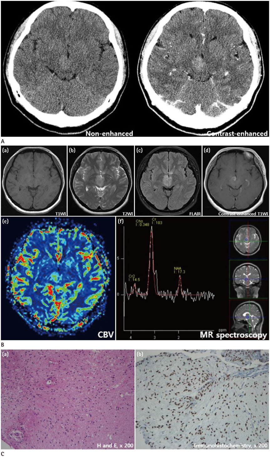
Fig. 1 Diffuse midline glioma, H3 K27M-mutant in the bilateral thalamus and midbrain in a 37-year-old woman, presenting with dizziness. A. Axial non-enhanced computed tomography scan revealing an ill-defined hyper-attenuated mass in the right thalamus. B. (a) Axial pre-contrast T1WI revealing a homogeneously hypointense lesion in the right thalamus. (b) Axial T2WI demonstrating a hyperintense lesion in the bilateral thalamus. (c) Axial FLAIR image revealing a hyperintense lesion in the bilateral thalamus. (d) Axial post-contrast T1WI revealing a focal irregular enhancement in the right thalamus. (e) CBV parameter map revealing an increased signal in the right thalamus. (f) MR spectroscopy: an increased choline peak and a markedly decreased N-acetylaspartate peak are apparent in the right thalamus. CBV = cerebral blood volume, FLAIR = fluid-attenuated inversion recovery, MR = magnetic resonance, T1WI = T1-weighted image, T2WI = T2-weighted image C. Photomicrograph: (a) hematoxylin and eosin stain, demonstrating dense cellularity and occasional nuclear atypia. (b) Immunohistochemistry for histone H3 K27M-mutant protein revealing the nuclei of a majority of tumor cells (brown stain) with only few normal, residual cells (blue) (original magnification × 200).
Reference
-
1. Louis DN, Perry A, Reifenberger G, von Deimling A, Figarella-Branger D, Cavenee WK, et al. The 2016 World Health Organization classification of tumors of the central nervous system: a summary. Acta Neuropathol. 2016; 131:803–820.2. Solomon DA, Wood MD, Tihan T, Bollen AW, Gupta N, Phillips JJ, et al. Diffuse midline gliomas with histone H3-K27M mutation: a series of 47 cases assessing the spectrum of morphologic variation and associated genetic alterations. Brain Pathol. 2016; 26:569–580.3. Aboian MS, Solomon DA, Felton E, Mabray MC, Villanueva-Meyer JE, Mueller S, et al. Imaging characteristics of pediatric diffuse midline gliomas with histone H3 K27M mutation. AJNR Am J Neuroradiol. 2017; 38:795–800.4. Guillamo JS, Monjour A, Taillandier L, Devaux B, Varlet P, Haie-Meder C, et al. Brainstem gliomas in adults: prognostic factors and classification. Brain. 2001; 124(Pt 12):2528–2539.5. Balasa A, Balasa R, Egyed-Zsigmond I, Chinezu R. Bilateral thalamic glioma: case report and review of the literature. Turk Neurosurg. 2016; 26:321–324.6. López G, Oberheim Bush NA, Berger MS, Perry A, Solomon DA. Diffuse non-midline glioma with H3F3A K27M mutation: a prognostic and treatment dilemma. Acta Neuropathol Commun. 2017; 5:38.7. Lakhan SE, Harle L. Difficult diagnosis of brainstem glioblastoma multiforme in a woman: a case report and review of the literature. J Med Case Rep. 2009; 3:87.8. Fischbein NJ, Prados MD, Wara W, Russo C, Edwards MS, Barkovich AJ. Radiologic classification of brain stem tumors: correlation of magnetic resonance imaging appearance with clinical outcome. Pediatr Neurosurg. 1996; 24:9–23.9. Osborn AG. Astrocytomas. In : Osborn AG, editor. Osborn's brain: imaging, pathology, and anatomy. 1st ed. Salt Lake City, UT: Amirsys;2012. p. 453–492.10. Rachinger W, Grau S, Holtmannspötter M, Herms J, Tonn JC, Kreth FW. Serial stereotactic biopsy of brainstem lesions in adults improves diagnostic accuracy compared with MRI only. J Neurol Neurosurg Psychiatry. 2009; 80:1134–1139.
- Full Text Links
-
- Actions
-
Cited
- CITED
-
- Close
- Share
-
- Similar articles
-
- Clinical Features and Prognosis of Diffuse Midline Glioma: A Series of 24 Cases
- Four Cases of Paramedian Thalamopeduncular Artery Infarction
- Astrocytoma in the Bilateral Thalamus: A Case Report
- Epigenetic and Metabolic Changes in Diffuse Intrinsic Pontine Glioma
- A Case of Bilateral Oculomotor Nuclear Palsy
