Extra-Axial and Clear Cell Type Ependymoma, Mimicking a Convexity Meningioma
- Affiliations
-
- 1Department of Neurosurgery, Seoul St. Mary's Hospital, College of Medicine, The Catholic University of Korea, Seoul, Koera. ssjeun@catholic.ac.kr
- 2Department of Hospital Pathology, Seoul St. Mary's Hospital, College of Medicine, The Catholic University of Korea, Seoul, Koera.
- KMID: 2396459
- DOI: http://doi.org/10.14791/btrt.2017.5.2.127
Abstract
- A 33-year-old woman presented with tingling and paresthesia on left extremity for 2 months. Magnetic resonance imaging revealed that the tumor was iso- and hypo-intensity on T1-weighted image, mixed iso- and high-signal intensity on T2-weighted images and heterogeneously enhanced with rim enhancement. Neither arachnoid cleft nor dural tail was certain but mass was located extra-axially so meningioma was suspected. During operation, tumor wasn't attached to dura at all but arachnoid attachment was seen. Pathologically, clear cell type ependymoma was confirmed. Details of diagnosis and treatment of this tumor is described.
Keyword
MeSH Terms
Figure
-
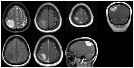
Fig. 1 A contrast-enhanced magnetic resonance imaging showed 41×42×29 mm sized extra-axial and heterogeneously enhanced mass on right parietal convexity, which seems to be originated from meninges, but dural-tail sign wasn't significant.
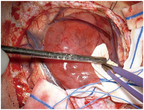
Fig. 2 Operative findings of extra-axial ependymoma. Unlike meningioma, mass was completely separated from dura mater without bone invasion. The mass was vascular, reddish, rubbery in consistency, and extra-axial with focal adhesion to arachnoid mater.
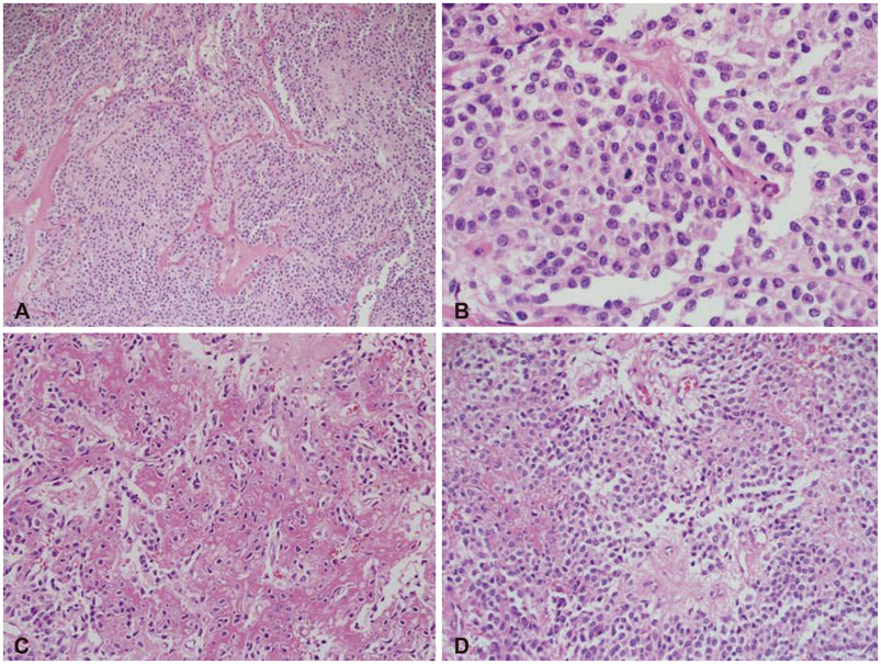
Fig. 3 Histopathologic findings stained hematoxylin and eosin, showed monomorphic cells with high cell density and delicate capillary blood vessels (×40) (A), clear perinuclear halo due to cytoplasmic clearing and bland cytology and mitotic count was 7 per 10 high power fields in the mostly active area (×400) (B). There were multifocal necrotic areas, microvascular proliferation and hyalinizing areas were noted in the part of tumor (×100) (C) and perivascular pseudorosettes found vaguely in the focal area (×100) (D).
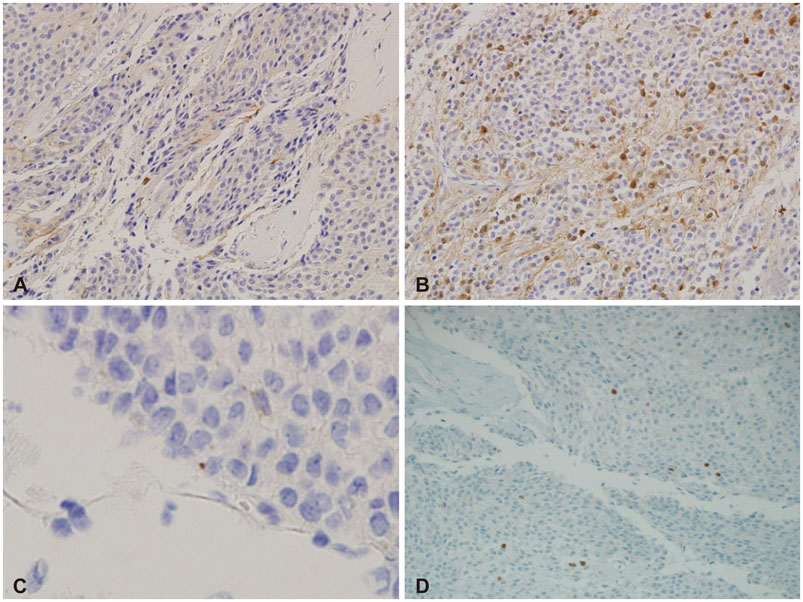
Fig. 4 Immunohistochemistry findings showed weakly positive GFAP (×100) (A), S-100 protein multifocally expressed (×100) (B), EMA manifested as dot-like perinuclear cytoplasmic structures of a few tumor cells (×400) (C), and sparse OLIG2 expression (×100) (D).
Reference
-
1. Youkilis AS, Park P, McKeever PE, Chandler WF. Parasagittal ependymoma resembling falcine meningioma. AJNR Am J Neuroradiol. 2001; 22:1105–1108.2. Mermuys K, Jeuris W, Vanhoenacker PK, Van Hoe L, D'Haenens P. Best cases from the AFIP: supratentorial ependymoma. Radiographics. 2005; 25:486–490.3. Fouladi M, Helton K, Dalton J, et al. Clear cell ependymoma: a clinicopathologic and radiographic analysis of 10 patients. Cancer. 2003; 98:2232–2244.
Article4. Hayashi K, Tamura M, Shimozuru T, et al. Extra-axial ependymoma–case report. Neurol Med Chir (Tokyo). 1994; 34:295–299.5. Koeller KK, Sandberg GD. Armed Forces Institute of Pathology. From the archives of the AFIP. Cerebral intraventricular neoplasms: radiologic-pathologic correlation. Radiographics. 2002; 22:1473–1505.6. Roncaroli F, Consales A, Fioravanti A, Cenacchi G. Supratentorial cortical ependymoma: report of three cases. Neurosurgery. 2005; 57:E192. discussion E192.
Article7. Oya N, Shibamoto Y, Nagata Y, Negoro Y, Hiraoka M. Postoperative radiotherapy for intracranial ependymoma: analysis of prognostic factors and patterns of failure. J Neurooncol. 2002; 56:87–94.8. Mansur DB, Perry A, Rajaram V, et al. Postoperative radiation therapy for grade II and III intracranial ependymoma. Int J Radiat Oncol Biol Phys. 2005; 61:387–391.
Article9. Wani NA, Mir F, Qayum A, Nazir P. Sacrococcygeal extraspinal ependymoma. Acta Neurochir (Wien). 2010; 152:917–918.
Article10. Ma YT, Ramachandra P, Spooner D. Case report: primary subcutaneous sacrococcygeal ependymoma: a case report and review of the literature. Br J Radiol. 2006; 79:445–447.
Article
- Full Text Links
-
- Actions
-
Cited
- CITED
-
- Close
- Share
-
- Similar articles
-
- Supratentorial Anaplastic Ependymoma Mimicking an Extra-Axial Tumor: A Case Report
- Metaplastic Meningioma Overspreading the Cerebral Convexity
- A Case of Supratentorial Intra-axial Ependymoma Showing Exophytic Growth
- A Case of Extra-axial Anaplastic Meningioma with Direct Orbital Extension
- Thoracic Intramedullay Clear Cell Meningioma
