Supratentorial Pilocytic Astrocytoma Mimicking Convexity Meningioma with Early Anaplastic Transformation: A Case Report
- Affiliations
-
- 1Department of Neurosurgery, Pusan National University Hospital, Busan, Korea. chwachoi@pusan.ac.kr
- 2Department of Neurosurgery, Pusan National University Yangsan Hospital, Yangsan, Korea.
- KMID: 2396455
- DOI: http://doi.org/10.14791/btrt.2017.5.2.105
Abstract
- Meningiomas and pilocytic astrocytomas are benign intracranial tumors. Pilocytic astrocytomas arises frequently at the posterior fossa in childhood. Meningiomas have several image findings, such as a dural tail sign, bony erosion, and sunburst appearance on angiography. However, pilocytic astrocytomas with these findings have been rarely reported. In this report, we describe a mass with typical image findings of a meningioma, but diagnosed as a supratentorial pilocytic astrocytoma with early anaplastic transformation.
Keyword
Figure
-

Fig. 1 Preoperative radiographic images. A: T2 weighted MRI showing a mass with multiple cystic portion and peritumoral edema. B: MRI with gadolinium reveals a well-enhancing mass with large dural base on the right frontal convexity area. C: Dura tail signs (black arrows) are identified on MRI with gadolinium. D: Bony thinning on right frontal bone (white arrow) is revealed on computed tomography scans. E: The angiographic image demonstrates sunburst appearance (dotted arrow). F: Sunburst appearance also shows on the lateral image (dotted arrow). MRI, magnetic resonance imaging.
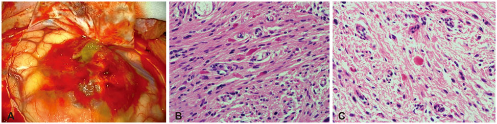
Fig. 2 Intraoperative photography (A) reveals a non-dural based and cystic mass. Photomicrograph of tumor shows Rosenthal fibers (B) and eosinophilic granular bodies (hematoxylin-eosin stain, ×200) (C).
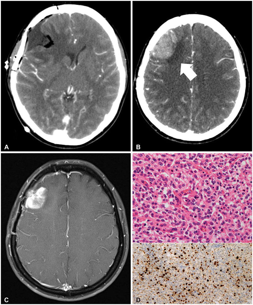
Fig. 3 Postoperative radiographic and pathologic findings. A: No remnant mass is identified on postoperative enhanced CT scans. B: Follow-up CT scans after 9 months shows a mass lesion suspicious of tumor recurrence (white arrow). C: Magnetic resonance imaging with gadolinium reveals a new well-enhancing mass on previous operative site. D: Pathologic findings shows increased cellularity, increased nuclear pleomorphism, and some mitoses (3 mitoses/10 high power field). There was no necrosis. Ki-index was about 30% (hematoxylin-eosin stain, ×200). CT, computed tomography.
Reference
-
1. Skipworth JR, Hill CS, Jones T, Foster J, Chopra I, Powell M. Pilocytic astrocytoma mimicking craniopharyngioma: a case series. Ann R Coll Surg Engl. 2012; 94:e125–e128.
Article2. Hong CS, Lehman NL, Sauvageau E. A pilocytic astrocytoma mimicking a clinoidal meningioma. Case Rep Radiol. 2014; 2014:524574.
Article3. Shibahara I, Kawaguchi T, Kanamori M, et al. Pilocytic astrocytoma with histological malignant features without previous radiation therapy--case report. Neurol Med Chir (Tokyo). 2011; 51:144–147.
Article4. Qi ST, Liu Y, Pan J, Chotai S, Fang LX. A radiopathological classification of dural tail sign of meningiomas. J Neurosurg. 2012; 117:645–653.
Article5. Watts J, Box G, Galvin A, Brotchie P, Trost N, Sutherland T. Magnetic resonance imaging of meningiomas: a pictorial review. Insights Imaging. 2014; 5:113–122.
Article6. Guan TK, Pancharatnam D, Chandran H, Hooi TK, Kumar G, Ganesan D. Infratentorial benign cystic meningioma mimicking a hemangioblastoma radiologically and a pilocytic astrocytoma intraoperatively: a case report. J Med Case Rep. 2013; 7:87.
Article7. Fortuna A, Ferrante L, Acqui M, Guglielmi G, Mastronardi L. Cystic meningiomas. Acta Neurochir (Wien). 1988; 90:23–30.
Article8. Chourmouzi D, Papadopoulou E, Konstantinidis M, et al. Manifestations of pilocytic astrocytoma: a pictorial review. Insights Imaging. 2014; 5:387–402.
Article9. Brown PD, Buckner JC, O'Fallon JR, et al. Adult patients with supratentorial pilocytic astrocytomas: a prospective multicenter clinical trial. Int J Radiat Oncol Biol Phys. 2004; 58:1153–1160.
Article10. Krieger MD, Gonzalez-Gomez I, Levy ML, McComb JG. Recurrence patterns and anaplastic change in a long-term study of pilocytic astrocytomas. Pediatr Neurosurg. 1997; 27:1–11.
Article11. Otero-Rodríguez A, Sarabia-Herrero R, García-Tejeiro M, Zamora-Martínez T. Spontaneous malignant transformation of a supratentorial pilocytic astrocytoma. Neurocirugia (Astur). 2010; 21:245–252.
Article12. Louis DN, Ohgaki H, Wiestler OD, Cavenee WK. WHO Classification of Tumours of the Central Nervous System, Revised. 4th ed. France: International Agency for Research on Cancer;2016.13. Rodriguez FJ, Scheithauer BW, Burger PC, Jenkins S, Giannini C. Anaplasia in pilocytic astrocytoma predicts aggressive behavior. Am J Surg Pathol. 2010; 34:147–160.
Article
- Full Text Links
-
- Actions
-
Cited
- CITED
-
- Close
- Share
-
- Similar articles
-
- Pilocytic Astrocytoma Occuring in a Patient Treated with Gamma Knife Surgery: Case Report
- Collision Tumor of Meningioma and Anaplastic Astrocytoma
- Multiple Solid Pilocytic Astrocytomas in Cerebellum with Neurofibromatosis Type I: A Case Report
- Anaplastic Cystic Meningioma
- CT and MR Findings of Supratentorial Pilocytic Astrocytoma
