J Korean Med Sci.
2017 Dec;32(12):2069-2072. 10.3346/jkms.2017.32.12.2069.
A Case of Multiple Cardiovascular and Tracheal Anomalies Presented with Wolff-Parkinson-White Syndrome in a Middle-aged Adult
- Affiliations
-
- 1Department of Internal Medicine, Hallym University Kangdong Sacred Heart Hospital, Seoul, Korea.
- 2Department of Internal Medicine, Jeju National University School of Medicine, Jeju National University Hospital, Jeju, Korea. masque70@dreamwiz.com
- 3Department of Radiology, Jeju National University School of Medicine, Jeju National University Hospital, Jeju, Korea.
- KMID: 2396388
- DOI: http://doi.org/10.3346/jkms.2017.32.12.2069
Abstract
- Congenital cardiovascular anomalies, such as dextrocardia, persistent left superior vena cava (SVC), and pulmonary artery (PA) sling, are rare disorders. These congenital anomalies can occur alone, or coincide with other congenital malformations. In the majority of cases, congenital anomalies are detected early in life by certain signs and symptoms. A 56-year-old man with no previous medical history was admitted due to recurrent wide QRS complex tachycardia with hemodynamic collapse. A chest radiograph showed dextrocardia. After synchronized cardioversion, an electrocardiogram revealed Wolff-Parkinson-White (WPW) syndrome. Persistent left SVC, PA sling, and right tracheal bronchus were also detected by a chest computed tomography (CT) scan. He was diagnosed with paroxysmal supraventricular tachycardia (PSVT) associated with WPW syndrome, and underwent radiofrequency ablation. We reported the first case of situs solitus dextrocardia coexisting with persistent left SVC, PA sling and right tracheal bronchus presented with WPW and PSVT in a middle-aged adult. In patients with a cardiovascular anomaly, clinicians should consider thorough evaluation of possibly combined cardiovascular and airway malformations and cardiac dysrhythmia.
Keyword
MeSH Terms
Figure
-
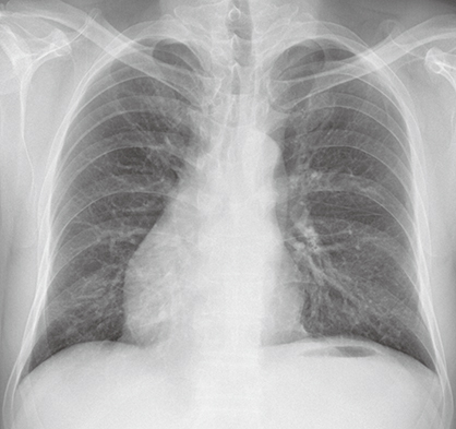
Fig. 1 The initial chest X-ray, showing dextrocardia.
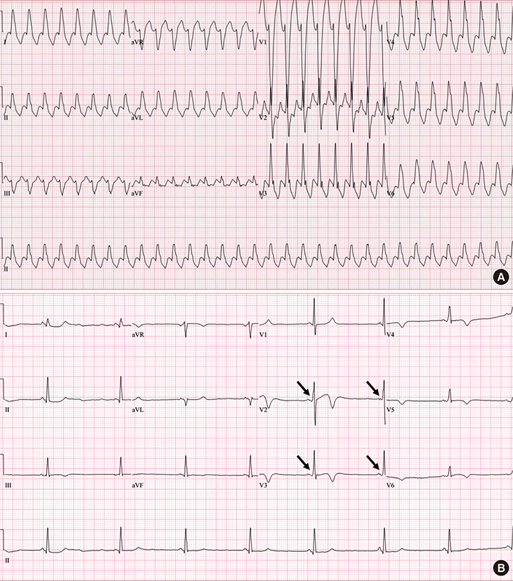
Fig. 2 Electrocardiograms. (A) An initial 12-lead electrocardiogram showed a heart rate of 190 bpm and wide QRS complex tachycardia. (B) After administration of intravenous adenosine, a follow-up electrocardiogram revealed bradycardia with a shortened PR interval and a slurring and slow rise of the initial upstroke of the QRS complex (black arrows).
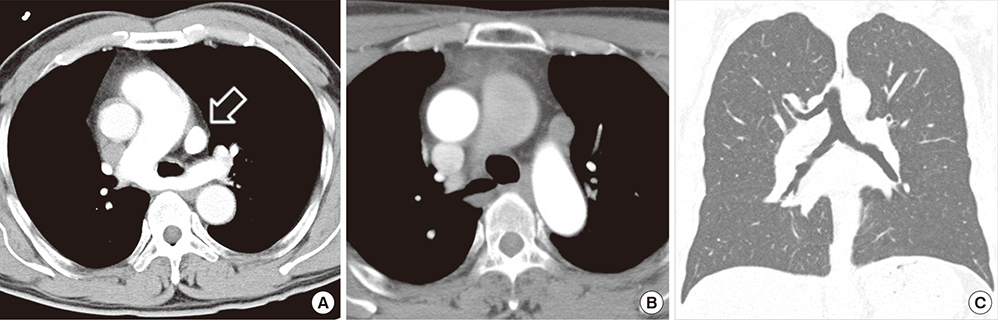
Fig. 3 Chest CT images. (A) An axial chest CT image reveals persistent left SVC (arrow) and PA sling. (B, C) Both axial and coronal chest CT images reveal right tracheal bronchus. CT = computed tomography, SVC = superior vena cava, PA = pulmonary artery.
Reference
-
1. Maldjian PD, Saric M. Approach to dextrocardia in adults [review]. AJR Am J Roentgenol. 2007; 188:S39–S49.2. Bohun CM, Potts JE, Casey BM, Sandor GG. A population-based study of cardiac malformations and outcomes associated with dextrocardia. Am J Cardiol. 2007; 100:305–309.3. Pálinkás A, Nagy E, Forster T, Morvai Z, Nagy E, Varga A. A case of absent right and persistent left superior vena cava. Cardiovasc Ultrasound. 2006; 4:6.4. Gonzalez-Juanatey C, Testa A, Vidan J, Izquierdo R, Garcia-Castelo A, Daniel C, Armesto V. Persistent left superior vena cava draining into the coronary sinus: report of 10 cases and literature review. Clin Cardiol. 2004; 27:515–518.5. Morgan LG, Gardner J, Calkins J. The incidental finding of a persistent left superior vena cava: implications for primary care providers-case and review. Case Rep Med. 2015; 2015:198754.6. Doig JC, Saito J, Harris L, Downar E. Coronary sinus morphology in patients with atrioventricular junctional reentry tachycardia and other supraventricular tachyarrhythmias. Circulation. 1995; 92:436–441.7. Morgan DR, Hanratty CG, Dixon LJ, Trimble M, O’Keeffe DB. Anomalies of cardiac venous drainage associated with abnormalities of cardiac conduction system. Europace. 2002; 4:281–287.8. Lee KJ, Kamagata S, Hirobe S, Toma M, Furukawa T, Fukushima N, Inomata Y. Aortopexy for tracheomalacia with dextrocardia, pulmonary artery sling, and congenital tracheal stenosis. Ann Thorac Surg. 2009; 88:1345–1348.9. Chen SJ, Lee WJ, Lin MT, Wang JK, Chang CI, Chiu IS, Wu MH. Left pulmonary artery sling complex: computed tomography and hypothesis of embryogenesis. Ann Thorac Surg. 2007; 84:1645–1650.10. Dupuis C, Vaksmann G, Pernot C, Gerard R, Martinez J, Van Egmond H. Asymptomatic form of left pulmonary artery sling. Am J Cardiol. 1988; 61:177–181.11. Shon TS, Moon KW, Yoo KD, Youn HJ, So SC, Kwak KK, Park HK, Chung WS, Han SK, Hong SJ. Anomalous origin of left coronary artery from pulmonary artery: report of an adult case. Korean Circ J. 1999; 29:528–531.12. Kim JG, Youn HJ, Kim GH, Park MH, Hur J, Yu JS, Jung SY, An SH. Incidentally detected situs ambiguous in adults. J Cardiovasc Ultrasound. 2011; 19:211–215.
- Full Text Links
-
- Actions
-
Cited
- CITED
-
- Close
- Share
-
- Similar articles
-
- One Case of Cerebral Embolism Associated with Paroxysmal Tachycardia in Wolff-Parkinson-White Syndrome
- Two Cases of Wolff-Parkinson-White Syndrome in a Family
- Wolff-Parkinson-White Syndrome Treated with Radiofrequency Ablation in Father and His Son
- Wolff-Parkinson-White Syndrome in a Patient With Mitochondrial Encephalopathy, Lactic Acidosis and Stroke-Like Episodes Syndrome
- A Case of Early Onset MELAS Patient with Wolff-Parkinson-White Syndrome
