Ann Dermatol.
2017 Dec;29(6):806-808. 10.5021/ad.2017.29.6.806.
A Case of Segmental (Zosteriform) Juvenile Xanthogranuloma
- Affiliations
-
- 1Department of Dermatology, Incheon St. Mary's Hospital, College of Medicine, The Catholic University of Korea, Incheon, Korea. hazelkimhoho@gmail.com
- KMID: 2395197
- DOI: http://doi.org/10.5021/ad.2017.29.6.806
Abstract
- No abstract available.
MeSH Terms
Figure
-
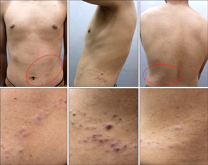
Fig. 1 Multiple, variably-sized, brown to skin-colored nodules along the waist (dotted circles).

Fig. 2 (A, B, D) Histopathologic examination shows dense lymphohistiocytic infiltration and toutontype giant cells with foamy cytoplasm in the dermis. (C) Histiocytic cells are positive in CD68 stain.
Reference
-
1. Garay M, Moreno S, Apreéa G, Pizzi-Parra N. Linear juvenile xanthogranuloma. Pediatr Dermatol. 2004; 21:513–515.
Article2. Kaur MR, Brundler MA, Stevenson O, Moss C. Disseminated clustered juvenile xanthogranuloma: an unusual morphological variant of a common condition. Clin Exp Dermatol. 2008; 33:575–577.
Article3. Ng SY. Segmental juvenile xanthogranuloma. Pediatr Dermatol. 2014; 31:615–617.
Article4. Kiorpelidou D, Stergiopoulou C, Zioga A, Bassukas ID. Linear-agminated juvenile xanthogranulomas. Int J Dermatol. 2008; 47:387–389.
Article5. Soon SL, Howard AK, Washington CV. Multiple, clustered dermatofibroma: a rare clinical variant of dermatofibroma. J Cutan Med Surg. 2003; 7:455–457.
Article
