Ann Dermatol.
2017 Apr;29(2):258-260. 10.5021/ad.2017.29.2.258.
A Case of Exophytic Pilomatricoma Clinically Resembling Keratoacanthoma
- Affiliations
-
- 1Department of Dermatology, Korea University Ansan Hospital, College of Medicine, Korea University, Ansan, Korea. kumcihk@korea.ac.kr
- KMID: 2394863
- DOI: http://doi.org/10.5021/ad.2017.29.2.258
Abstract
- No abstract available.
MeSH Terms
Figure
-
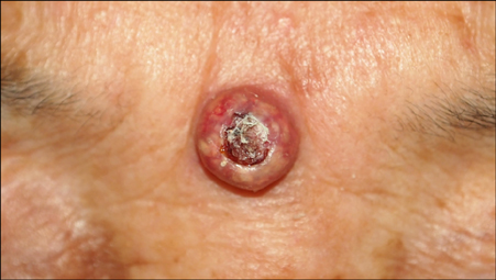
Fig. 1 Dome-shaped mass in the glabella with brownish crust in the central portion. Mixture of erythematous and whitish color can be observed in the peripheral area.
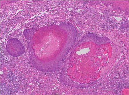
Fig. 2 Multiple masses within dermis. Eosinophilic ghost cells can be seen in the center and basaloid cells in the peripheral portion (H&E, ×40).
Reference
-
1. Holme SA, Varma S, Holt PJ. The first case of exophytic pilomatricoma in an Asian male. Pediatr Dermatol. 2001; 18:498–500.
Article2. Pirouzmanesh A, Reinisch JF, Gonzalez-Gomez I, Smith EM, Meara JG. Pilomatrixoma: a review of 346 cases. Plast Reconstr Surg. 2003; 112:1784–1789.3. Faust HB, Clark RE, Kamino H. Hyperkeratotic nodule. Keratoacanthomalike pilomatricoma. Arch Dermatol. 1996; 132:573. 576.4. Kang HY, Kang WH. Guess what! Perforating pilomatricoma resembling keratoacanthoma. Eur J Dermatol. 2000; 10:63–64.5. Kost DM, Smart DR, Jones WB, Bain M. A perforating pilomatricomal horn on the arm of an 11-year-old girl. Dermatol Online J. 2014; 20:22371.
Article
- Full Text Links
-
- Actions
-
Cited
- CITED
-
- Close
- Share
-
- Similar articles
-
- A Case of Exophytic Pilomatricoma with Perforating Figure
- A Case of Keratoacanthoma on the Lower Lip
- Pigmented Pilomatricoma on the Ear Resembling Vascular Tumor before Surgery: A Case Report
- Multiple Giant Keratoacanthoma Treated with Acitretin
- Topical Treatment with 5% Imiquimod for Solitary Keratoacanthoma
