Clinical Outcome of Magnetic Resonance Imaging-Detected Additional Lesions in Breast Cancer Patients
- Affiliations
-
- 1Department of Surgery and Institute for Medical Sciences, Chonbuk National University Medical School, Jeonju, Korea. yhj0903@jbnu.ac.kr
Abstract
- PURPOSE
The aim of this study was to investigate the clinical outcome of additional breast lesions identified with breast magnetic resonance imaging (MRI) in breast cancer patients.
METHODS
A total of 153 patients who underwent breast MRI between July 2006 and March 2008 were retrospectively reviewed. Thirty-three patients (21.6&) were recommended for second-look ultrasound (US) for further characterization of additional lesions detected on breast MRI and these patients constituted our study population.
RESULTS
Assessment for lesions detected on breast MRI consisted of the following: 25 benign lesions (73.5&), two indeterminate (5.9%), and seven malignant (20.6%) in 33 patients. Second-look US identified 12 additional lesions in 34 lesions (35.3%) and these lesions were confirmed by histological examination. Of the 12 lesions found in the 11 patients, six (50.0%) including one contralateral breast cancer were malignant. The surgical plan was altered in 18.2% (six of 33) of the patients. The use of breast MRI justified a change in treatment for four patients (66.7%) and caused two patients (33.3&) to undergo unwarranted additional surgical procedures.
CONCLUSION
Breast MRI identified additional multifocal or contralateral cancer which was not detected initially on conventional imaging in breast cancer patients. Breast MRI has become an indispensable modality in conjunction with conventional modalities for preoperative evaluation of patients with operable breast cancer.
MeSH Terms
Figure
-

Figure 1 Assessment and pathologic results of additional lesions on magnetic resonance imaging (MRI). Additional 34 lesions on breast MRI were found in 33 patients. Twenty-two lesions which were assessed as benign lesion on breast MRI were not identified the additional lesions on second-look ultrasound (US). Twelve additional lesions in 11 patients have been diagnosed as follows. *One patient has two malignant assessed lesions in each breast.
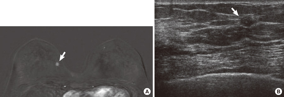
Figure 2 A 59-year-old woman with known right breast cancer (4-o'clock position) who underwent preoperative breast magnetic resonance imaging (MRI). (A) Subtraction axial MR image shows unexpected oval shaped enhancing lesion (arrow) at 3-o'clock position in the right breast. It was classified as malignant. (B) On second-look ultrasound, 0.7 cm sized, oval, ill defined, and hypoechoic lesion was found at the identical location. Sonographic findings suggest BI-RADS 4C and pathologic result was invasive ductal carcinoma.
Reference
-
1. Kuhl CK. MRI of breast tumors. Eur Radiol. 2000. 10:46–58.
Article2. Braun M, Pölcher M, Schrading S, Zivanovic O, Kowalski T, Flucke U, et al. Influence of preoperative MRI on the surgical management of patients with operable breast cancer. Breast Cancer Res Treat. 2008. 111:179–187.
Article3. Linda A, Zuiani C, Londero V, Bazzocchi M. Outcome of initially only magnetic resonance mammography-detected findings with and without correlate at second-look sonography: distribution according to patient history of breast cancer and lesion size. Breast. 2008. 17:51–57.
Article4. Wiratkapun C, Duke D, Nordmann AS, Lertsithichai P, Narra V, Barton PT, et al. Indeterminate or suspicious breast lesions detected initially with MR imaging: value of MRI-directed breast ultrasound. Acad Radiol. 2008. 15:618–625.
Article5. Houssami N, Hayes DF. Review of preoperative magnetic resonance imaging (MRI) in breast cancer: should MRI be performed on all women with newly diagnosed, early stage breast cancer? CA Cancer J Clin. 2009. 59:290–302.
Article6. Pengel KE, Loo CE, Teertstra HJ, Muller SH, Wesseling J, Peterse JL, et al. The impact of preoperative MRI on breast-conserving surgery of invasive cancer: a comparative cohort study. Breast Cancer Res Treat. 2009. 116:161–169.
Article7. LaTrenta LR, Menell JH, Morris EA, Abramson AF, Dershaw DD, Liberman L. Breast lesions detected with MR imaging: utility and histopathologic importance of identification with US. Radiology. 2003. 227:856–861.
Article8. Warren RM, Thompson D, Pointon LJ, Hoff R, Gilbert FJ, Padhani AR, et al. Evaluation of a prospective scoring system designed for a multicenter breast MR imaging screening study. Radiology. 2006. 239:677–685.
Article9. Schnall MD, Blume J, Bluemke DA, DeAngelis GA, DeBruhl N, Harms S, et al. Diagnostic architectural and dynamic features at breast MR imaging: multicenter study. Radiology. 2006. 238:42–53.
Article10. Abe H, Schmidt RA, Shah RN, Shimauchi A, Kulkarni K, Sennett CA, et al. MR-directed ("Second-Look") ultrasound examination for breast lesions detected initially on MRI: MR and sonographic findings. AJR Am J Roentgenol. 2010. 194:370–377.
Article11. Destounis S, Arieno A, Somerville PA, Seifert PJ, Murphy P, Morgan R, et al. Community-based practice experience of unsuspected breast magnetic resonance imaging abnormalities evaluated with second-look sonography. J Ultrasound Med. 2009. 28:1337–1346.
Article12. Tardivon AA, Athanasiou A, Thibault F, El Khoury C. Breast imaging and reporting data system (BIRADS): magnetic resonance imaging. Eur J Radiol. 2007. 61:212–215.
Article13. Liberman L, Morris EA, Dershaw DD, Abramson AF, Tan LK. MR imaging of the ipsilateral breast in women with percutaneously proven breast cancer. AJR Am J Roentgenol. 2003. 180:901–910.
Article14. Hollingsworth AB, Stough RG, O'Dell CA, Brekke CE. Breast magnetic resonance imaging for preoperative locoregional staging. Am J Surg. 2008. 196:389–397.
Article15. Sotome K, Yamamoto Y, Hirano A, Takahara T, Hasegawa S, Nakamaru M, et al. The role of contrast enhanced MRI in the diagnosis of non-mass image-forming lesions on breast ultrasonography. Breast Cancer. 2007. 14:371–380.
Article16. Mann RM, Kuhl CK, Kinkel K, Boetes C. Breast MRI: guidelines from the European Society of Breast Imaging. Eur Radiol. 2008. 18:1307–1318.
Article17. Shin JH, Han BK, Choe YH, Ko K, Choi N. Targeted ultrasound for MR-detected lesions in breast cancer patients. Korean J Radiol. 2007. 8:475–483.
Article18. Beran L, Liang W, Nims T, Paquelet J, Sickle-Santanello B. Correlation of targeted ultrasound with magnetic resonance imaging abnormalities of the breast. Am J Surg. 2005. 190:592–594.
Article19. Sim LS, Hendriks JH, Bult P, Fook-Chong SM. US correlation for MRI-detected breast lesions in women with familial risk of breast cancer. Clin Radiol. 2005. 60:801–806.
Article20. Jin JW, Yom CK, Koo MY, Moon BI, Choi KJ, Choi HY. The role and significance of preoperative breast MRI in the setting of breast cancer. J Breast Cancer. 2008. 11:146–150.
Article21. Mann RM, Loo CE, Wobbes T, Bult P, Barentsz JO, Gilhuijs KG, et al. The impact of preoperative breast MRI on the re-excision rate in invasive lobular carcinoma of the breast. Breast Cancer Res Treat. 2010. 119:415–422.
Article22. Fischer U, Zachariae O, Baum F, von Heyden D, Funke M, Liersch T. The influence of preoperative MRI of the breasts on recurrence rate in patients with breast cancer. Eur Radiol. 2004. 14:1725–1731.
Article23. Hata T, Takahashi H, Watanabe K, Takahashi M, Taguchi K, Itoh T, et al. Magnetic resonance imaging for preoperative evaluation of breast cancer: a comparative study with mammography and ultrasonography. J Am Coll Surg. 2004. 198:190–197.
Article24. Turnbull L, Brown S, Harvey I, Olivier C, Drew P, Napp V, et al. Comparative effectiveness of MRI in breast cancer (COMICE) trial: a randomized controlled trial. Lancet. 2010. 375:563–571.
Article
- Full Text Links
-
- Actions
-
Cited
- CITED
-
- Close
- Share
-
- Similar articles
-
- Tips for finding magnetic resonance imaging-detected suspicious breast lesions using second-look ultrasonography: a pictorial essay
- Second-look ultrasonography for MRI-detected suspicious breast lesions in patients with breast cancer
- Low Rates of Additional Cancer Detection by Magnetic Resonance Imaging in Newly Diagnosed Breast Cancer Patients Who Undergo Preoperative Mammography and Ultrasonography
- Targeted Ultrasound for MR-Detected Lesions in Breast Cancer Patients
- Clinical Experience of 3T Breast MRI in Detecting the Additional Lesions in Breast Cancer Patients
