Obstet Gynecol Sci.
2017 Sep;60(5):494-497. 10.5468/ogs.2017.60.5.494.
A case report of angioleiomyoma of uterus
- Affiliations
-
- 1Department of Obstetrics and Gynecology, Soonchunhyang University Bucheon Hospital, Soonchunhyang University College of Medicine, Bucheon, Korea. guardc@schmc.ac.kr
- 2Department of Pathology, Soonchunhyang University Bucheon Hospital, Soonchunhyang University College of Medicine, Bucheon, Korea.
- KMID: 2393853
- DOI: http://doi.org/10.5468/ogs.2017.60.5.494
Abstract
- Angioleiomyoma (AL) is a very rare benign tumor that originates from smooth muscle cells and has thick walled vessels. It may be found throughout the body but more frequently occurs in the lower extremities and rarely develops in the head and other parts of the body. This paper presents a case report of giant AL detected in a 33-year-old woman who complained of severe anemia, menorrhagia, and palpable lower abdominal mass. The patient underwent myomectomy and was diagnosed with AL based on the pathological report of mass. The effective treatment for AL is either simple hysterectomy or angiomyomectomy depending on the patient's desire to preserve fertility and symptom.
Keyword
MeSH Terms
Figure
-
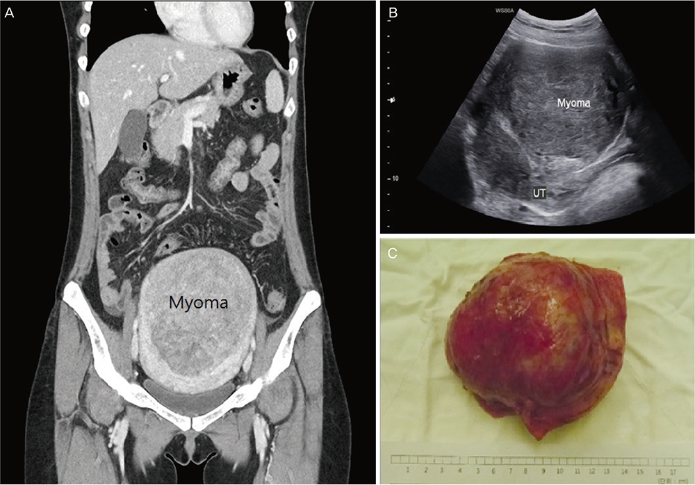
Fig. 1 (A) Computed tomography shows 14.2 cm huge uterine myoma with endometrial compression, (B) transabdominal ultrasound sonography shows huge myoma found anterior to the uterus, and (C) gross findings show a 12.5×9.0 cm2 sized gray firm mass after the operation. UT, uterus.

Fig. 2 (A) Microscopic findings indicate the tumor with interlacing short spindle cells and numerous thick walled vessels (hematoxylin and eosin stain, ×40), (B) immunohistochemical stain of smooth muscle actin (SMA) reveals diffuse positivity in the spindle tumor cells and swirling cells around the thick-walled blood vessels (immunohistochemistry of SMA, ×100).
Reference
-
1. Sharma C, Sharma M, Chander B, Soni A, Soni PK. Angioleiomyoma uterus in an adolescent girl: a highly unusual presentation. J Pediatr Adolesc Gynecol. 2014; 27:e69–e71.2. Hachisuga T, Hashimoto H, Enjoji M. Angioleiomyoma. A clinicopathologic reappraisal of 562 cases. Cancer. 1984; 54:126–130.3. Garg G, Mohanty SK. Uterine angioleiomyoma: a rare variant of uterine leiomyoma. Arch Pathol Lab Med. 2014; 138:1115–1118.4. Zizi-Sermpetzoglou A, Myoteri D, Arkoumani E, Koulia K, Tsavari A, Alamanou E, et al. Angioleiomyoma of the uterus: report of a distinctive benign leiomyoma variant. Eur J Gynaecol Oncol. 2015; 36:210–212.5. Handler M, Rezai F, Fless KG, Litinski M, Yodice PC. Uterine angioleiomyoma complicated by consumptive coagulopathy. Gynecol Oncol Case Rep. 2012; 2:89–91.6. Hsieh CH, Lui CC, Huang SC, Ou YC, ChangChien CC, Lan KC, et al. Multiple uterine angioleiomyomas in a woman presenting with severe menorrhagia. Gynecol Oncol. 2003; 90:348–352.7. Keerthi R, Nanjappa M, Deora SS, Kumaraswamy SV. Angioleiomyoma of cheek: report of two cases. J Maxillofac Oral Surg. 2009; 8:298–300.8. Ishikawa S, Fuyama S, Kobayashi T, Taira Y, Sugano A, Iino M. Angioleiomyoma of the tongue: a case report and review of the literature. Odontology. 2016; 104:119–122.
