Luteolin 5-O-glucoside from Korean Milk Thistle, Cirsium maackii, Exhibits Anti-Inflammatory Activity via Activation of the Nrf2/HO-1 Pathway
- Affiliations
-
- 1Department of Food Science and Human Nutrition, Chonbuk National University, Jeonju 54896, Republic of Korea.
- 2Department of Food and Life Science, Pukyong National University, Busan 48513, Republic of Korea. choijs@pknu.ac.kr
- 3Department of Pharmaceutical Engineering, Sangji University, Wonju 220-702, Republic of Korea.
- KMID: 2393799
- DOI: http://doi.org/10.20307/nps.2017.23.3.183
Abstract
- Luteolin 5-O-glucoside is the major flavonoid from Korean thistle, Cirsium maackii. We previously reported the anti-inflammatory activities of luteolin 5-O-glucoside in lipopolysaccharide (LPS)-stimulated RAW 264.7 cells. In this study, we determined the anti-inflammatory mechanisms of luteolin 5-O-glucoside through the inhibition of nitric oxide (NO) production in vitro and in vivo. Results revealed that luteolin 5-O-glucoside dose-dependently inhibited NO production and expression of iNOS and COX-2 in LPS-induced RAW 264.7 cells. Luteolin 5-O-glucoside also significantly inhibited the translocation of NF-κB, the activation of MAPKs, and ROS generation in LPS-induced RAW 264.7 cells. In addition, protein expressions of Nrf-2 and HO-1 were also upregulated by luteolin 5-O-glucoside treatment. Moreover, luteolin 5-O-glucoside inhibited λ-carrageenan-induced mouse paw edema by 65.34% and 48.31% at doses of 50 and 100 mg/kg body weight, respectively. These findings indicate potential anti-inflammatory effect of luteolin 5-O-glucoside particularly by downregulating NF-κB and upregulating HO-1/Nrf-2 pathway.
Keyword
MeSH Terms
Figure
-

Fig. 1 Cell viability of luteolin 5-O-glucoside measured by the MTT assay. Values represent the mean ± SD of three independent Experiments.

Fig. 2 Inhibitory effects of luteolin 5-O-glucoside on the production of NO in LPS-stimulated RAW 264.7 cells. Cells were pretreated with different concentrations of luteolin 5-O-glucoside for 2 h and then stimulated with LPS (1 µg/ml) for 24 h. The culture media were used to measure the amount of nitrite to determine NO production. Data are presented as mean ± SD of three independent experiments. #p < 0.05 indicates a significant difference from the control group. *p < 0.05 indicates a significant difference from the LPS-treated group.
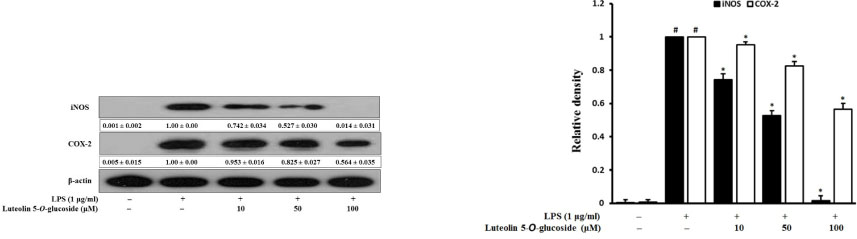
Fig. 3 Inhibitory effects of luteolin 5-O-glucoside on the expression of iNOS and COX-2 in LPS-stimulated RAW 264.7 cells. Cells were pretreated with the indicated concentration of luteolin 5-O-glucoside for 2 h and stimulated with LPS (1 µg/ml) for 18 h. The expression of iNOS, COX-2, and β-actin was detected by Western blotting using corresponding antibodies. The results presented are representative of three independent experiments. #p < 0.05 indicates a significant difference from the control group. *p < 0.05 indicates a significant difference from the LPS-treated group.

Fig. 4 Inhibitory effects of luteolin 5-O-glucoside on the expression of NF-κB in LPS-stimulated RAW 264.7 cells. Cells were pretreated with the indicated concentration of luteolin 5-O-glucoside for 2 h and stimulated with LPS (1 µg/ml) for 18 h. The expression of NF-κB and β-actin was detected by Western blot using the corresponding antibodies. The results presented are representative of three independent experiments. #p < 0.05 indicates a significant difference from the control group. *p < 0.05 indicates a significant difference from the LPS-treated group.

Fig. 5 Inhibitory effects of luteolin 5-O-glucoside on the expression of MAPKs in LPS-stimulated RAW 264.7 cells. Cells were pretreated with the indicated concentration of luteolin 5-O-glucoside for 2 h and stimulated with LPS (1 µg/ml) for 18 h. The expression of MAPKs was detected by Western blotting using corresponding antibodies. The results presented are representative of three independent experiments. #p < 0.05 indicates significant differences from the control group. *p < 0.05 indicates significant differences from the LPS-treated group.

Fig. 6 Inhibitory effects of luteolin 5-O-glucoside on the production of ROS in LPS-stimulated RAW 264.7 cells. Cells were pretreated with different concentration of luteolin 5-O-glucoside for 2 h and then stimulated with LPS (1 µg/ml) for 24 h. ROS levels were measured by fluorescence analysis of DCFH-DA. Data are presented as means ± SDs of three independent experiments. #p < 0.05 indicates a significant difference from the control group. *p < 0.05 indicates a significant difference from the LPS-treated group.

Fig. 7 Inhibitory effects of luteolin 5-O-glucoside on the expression of HO-1 and Nrf-2 in LPS-stimulated RAW 264.7 cells. Cells were pretreated with the indicated concentration of luteolin 5-O-glucoside for 2 h and stimulated with LPS (1 µg/ml) for 18 h. Expression of HO-1 and Nrf-2 was detected by Western blotting using the corresponding antibodies. The results presented are representative of three independent experiments. #p < 0.05 indicates a significant difference from the control group. *p < 0.05 indicates a significant difference from the LPS-treated group.

Fig. 8 Effects of luteolin 5-O-glucoside on carrageenan-induced hind paw edema in mice. Luteolin 5-O-glucoside was administered at doses of 25 mg/kg and 50 mg/kg 1 h before injection of carrageenan into the mice to alleviate acute inflammation. Paw volume was measured using a plethysmometer at 2 h, 5 h, and 7 h after carrageenan injection. The increase in paw volume was calculated based on the difference in volume between before carrageenan injection and at different time intervals after carrageenan injection. Values are expressed as the mean ± SD; n = 6 mice per group. *p < 0.05 indicates a significant difference from the vehicle control group. Indomethacin was used as a positive control.
Reference
-
1. Choudhari AS, Raina P, Deshpande MM, Wali AG, Zanwar A, Bodhankar SL, Kaul-Ghanekar R. J Ethnopharmacol. 2013; 150:215–222.2. Joung EJ, Lee B, Gwon WG, Shin T, Jung BM, Yoon NY, Choi JS, Oh CW, Kim HR. Int Immunopharmacol. 2015; 29:693–700.3. Giuliani C, Napolitano G, Bucci I, Montani V, Monaco F. Clin Ter. 2001; 152:249–253.4. May MJ, Ghosh S. Immunol Today. 1998; 19:80–88.5. Tak PP, Firestein GS. J Clin Invest. 2001; 107:7–11.6. Kim AR, Lee MS, Shin TS, Hua H, Jang BC, Choi JS, Byun DS, Utsuki T, Ingram D, Kim HR. Toxicol In Vitro. 2011; 25:1789–1795.7. Pae HO, Chung HT. Immune Netw. 2009; 9:12–19.8. Chen TY, Sun HL, Yao HT, Lii CK, Chen HW, Chen PY, Li CC, Liu KL. Food Chem Toxicol. 2013; 55:257–264.9. Taha R, Blaise G. Funct Food Health Dis. 2014; 4:510–523.10. Kim JG. Illustrated Natural Drugs Encyclopedia. Korea: Namsandang;1984. p. 37.11. Iwashina T, Ito T, Ootani S. Ann Tsukuba Bot Gard. 1989; 8:15–19.12. Jung HA, Kim YS, Choi JS. Food Chem Toxicol. 2009; 47:2790–2797.13. Jung HA, Jin SE, Min BS, Kim BW, Choi JS. Food Chem Toxicol. 2012; 50:2171–2179.14. Morris CJ. Methods Mol Biol. 2003; 225:115–121.15. Sharif O, Bolshakov VN, Raines S, Newham P, Perkins ND. BMC Immunol. 2007; 8:1–17.
Article16. Lee CB. Flora of Korea. Korea: Hyangmoonsa;1979. p. 274.17. Lee SJ. Korean Folk Medicine. Korea: Seoul National University;1966. p. 145–146.18. Keiser K, Johnson CC, Tipton DA. J Endod. 2000; 26:288–291.19. Meda L, Cassatella MA, Szendrei GI, Otvos L Jr, Baron P, Villalba M, Ferrari D, Rossi F. Nature. 1995; 374:647–650.20. Dandona P, Chaudhuri A, Dhindsa S. Diabetes Care. 2010; 33:1686–1687.21. Marks-Konczalik J, Chu SC, Moss J. J Biol Chem. 1998; 273:22201–22208.22. Islam MN, Choi RJ, Jin SE, Kim YS, Ahn BR, Zhao D, Jung HA, Choi JS. J Ethnopharmacol. 2012; 144:175–181.23. Chen JJ, Huang WC, Chen CC. Mol Biol Cell. 2005; 16:5579–5591.24. Rajapakse N, Kim MM, Mendis E, Kim SK. Immunology. 2008; 123:348–357.25. Kaminska B. Biochim Biophys Acta. 2005; 1754:253–262.26. Hancock JT, Desikan R, Neill SJ. Biochem Soc Trans. 2001; 29:345–350.27. Choi SY, Hwang JH, Ko HC, Park JG, Kim SJ. J Ethnopharmacol. 2007; 113:149–155.28. Siomek A. Acta Biochim Pol. 2012; 59:323–331.29. Ryan KA, Smith MF Jr, Sanders MK, Ernst PB. Infect Immun. 2004; 72:2123–2130.30. Kim JH, Choo YY, Tae N, Min BS, Lee JH. Int Immunopharmacol. 2014; 22:420–426.31. Lee IS, Lim J, Gal J, Kang JC, Kim HJ, Kang BY, Choi HJ. Neurochem Int. 2011; 58:153–160.32. Paine A, Eiz-Vesper B, Blasczyk R, Immenschuh S. Biochem Pharmacol. 2010; 80:1895–1903.33. Lee MY, Lee JA, Seo CS, Ha H, Lee H, Son JK, Shin HK. Food Chem Toxicol. 2011; 49:1047–1055.34. Tsoyi K, Lee TY, Lee YS, Kim HJ, Seo HG, Lee JH, Chang KC. Mol Pharmacol. 2009; 76:173–182.35. Salvemini D, Wang ZQ, Bourdon DM, Stern MK, Currie MG, Manning PT. Eur J Pharmacol. 1996; 303:217–220.36. Rocha AC, Fernandes ES, Quintão NL, Campos MM, Calixto JB. Br J Pharmacol. 2006; 148:688–695.37. Yuan G, Wahlqvist ML, He G, Yang M, Li D. Asia Pac J Clin Nutr. 2006; 15:143–152.38. Camuesco D, Comalada M, Rodríguez-Cabezas ME, Nieto A, Lorente MD, Concha A, Zarzuelo A, Gálvez J. Br J Pharmacol. 2004; 143:908–918.39. Halliwell B, Zhao K, Whiteman M. Free Radic Res. 2000; 33:819–830.40. Comalada M, Ballester I, Bailón E, Sierra S, Xaus J, Gálvez J, de Medina FS, Zarzuelo A. Biochem Pharmacol. 2006; 72:1010–1021.
- Full Text Links
-
- Actions
-
Cited
- CITED
-
- Close
- Share
-
- Similar articles
-
- Isolation and Quantitative Analysis of BACE1 Inhibitory Compounds from Cirsium maackii Flower
- Luteolin and luteolin-7-O-glucoside inhibit lipopolysaccharide-induced inflammatory responses through modulation of NF-kappaB/AP-1/PI3K-Akt signaling cascades in RAW 264.7 cells
- Luteolin and luteolin-7-O-glucoside protect against acute liver injury through regulation of inflammatory mediators and antioxidative enzymes in GalN/LPS-induced hepatitic ICR mice
- Luteolin Promotes Apoptosis of Endometriotic Cells and Inhibits the Alternative Activation of Endometriosis-Associated Macrophages
- Fraxetin Induces Heme Oxygenase-1 Expression by Activation of Akt/Nrf2 or AMP-activated Protein Kinase α/Nrf2 Pathway in HaCaT Cells
