An Unruptured Anterior Communicating Artery Aneurysm Presenting with Left Homonymous Hemianopsia: A Case Report
- Affiliations
-
- 1Department of Neurosurgery, Chung-Ang University Hospital, Seoul, Korea. nsnam@cau.ac.kr
- KMID: 2393385
- DOI: http://doi.org/10.7461/jcen.2017.19.2.92
Abstract
- Unruptured cerebral aneurysms sometimes present with visual symptomsdue to compression of the visual pathways. However, until now, unruptured anterior communicating artery (ACoA) aneurysms presenting visual field defects have been extremely rare. The authors report the case of a 51-year-old woman who presented with left homonymous hemianopsia. Radiological findings demonstrated an ACoA aneurysm filled with thrombus, that was compressing the optic chiasm and post-chiasmal tract. The patient underwent clipping of the aneurysm, which resolved the visual field defect. In cases of visual field defects, an ACoA aneurysm should be included in the differential diagnosis.
MeSH Terms
Figure
-
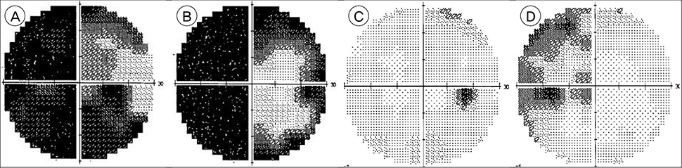
Fig. 1 Preoperative static visual field tests, right eye (A) and left eye (B), showing a left homonymous hemianopsia. Impovement of the visual fields in right eye (C) and left eye (D) at two months after surgery.
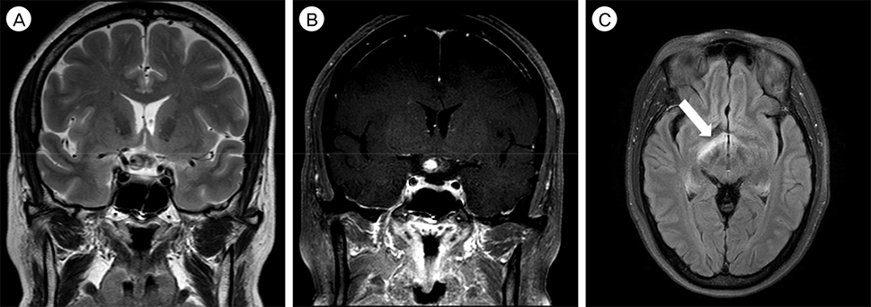
Fig. 2 (A, B) CoronalT2-weighted and T1-weighted with contrast images revealing a thrombosed aneurysm compressing the right side of the optic chiasm and postchiasmatic optic tract. (C) Axial T2-weighted FLAIR image showing the high signal intensity (arrow) of the right post-chiasmal optic tract.
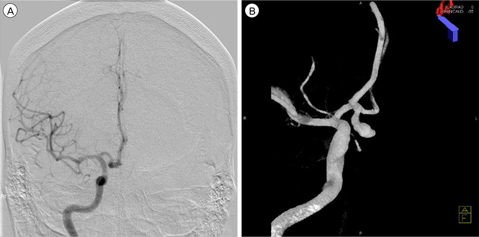
Fig. 3 Right internal carotid angiography, anteroposterior view (A) and 3D reconstruction (B), showing anterior communicating artery aneurysm projecting posteroinferiorly.
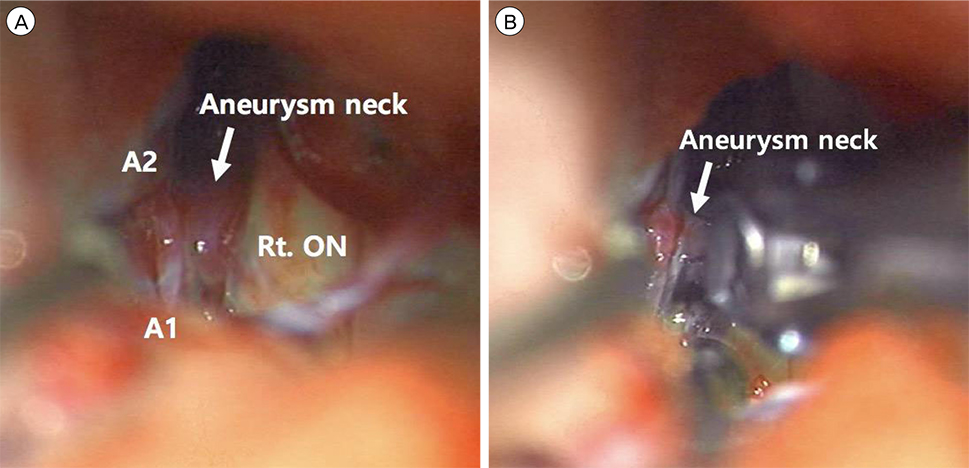
Fig. 4 Intraoperative photographs obtained before (A) and after (B) aneurysmal neck (arrow) clipping. ON = optic nerve.
Reference
-
1. Aoki N. Partially thrombosed aneurysm presenting as the sudden onset of bitemporal hemianopsia. Neurosurgery. 1988; 03. 22(3):564–566.
Article2. Carneiro A, Rane N, Küker W, Cellerini M, Corkill R, Byrne JV. Volume changes of extremely large and giant intracranial aneurysms after treatment with flow diverter stents. Neuroradiology. 2014; 01. 56(1):51–58.
Article3. Date I, Asari S, Ohmoto T. Cerebral aneurysms causing visual symptoms: their features and surgical outcome. Clin Neurol Neurosurg. 1998; 12. 100(4):259–267.
Article4. Ferguson GG, Drake CG. Carotid-ophthalmic aneurysms: visual abnormalities in 32 patients and the results of treatment. Surg Neurol. 1981; 07. 16(1):1–8.
Article5. Hagihara N, Abe T, Yoshioka F, Watanabe M, Tabuchi K. Photophobia as the visual manifestation of chiasmal compression by unruptured anterior communicating artery aneurysm. Case report. Neurol Med Chir (Tokyo). 2009; 04. 49(4):159–161.6. Höök O, Norlén G. Aneurysms of the anterior communicating artery. Acta Neurol Scand. 1964; 09. 40(3):219–240.
Article7. La Pira B, Brinjikji W, Hunt C, Chen JJ, Lanzino G. Reversible edema-like changes along the optic tract following Pipeline-assisted coiling of a large anterior communicating artery aneurysm. J Neuroophthalmol. 2017; 06. 37(2):154–158.
Article8. McAuliffe W, Wycoco V, Rice H, Phatouros C, Singh TJ, Wenderoth J. Immediate and midterm results following treatment of unruptured intracranial aneurysms with the pipeline embolization device. AJNR Am J Neuroradiol. 2012; 01. 33(1):164–170.
Article9. Moteki Y, Kawamoto T, Namioka T, Yokote A, Kawamata T. Progressive visual field defect caused by an unruptured middle cerebral artery aneurysm. Clin Neurol Neurosurg. 2013; 10. 115(10):2182–2185.
Article10. Norwood EG, Kline LB, Chandra-Sekar B, Harsh GR 3rd. Aneurysmal compression of the anterior visual pathways. Neurology. 1986; 08. 36(8):1035–1041.
Article11. Park JH, Park SK, Kim TH, Shin JJ, Shin HS, Hwang YS. Anterior communicating artery aneurysm related to visual symptoms. J Korean Neurosurg Soc. 2009; 09. 46(3):232–238.
Article12. Peiris JB, Ross Russell RW. Giant aneurysms of the carotid system presenting as visual field defect. J Neurol Neurosurg Psychiatry. 1980; 12. 43(12):1053–1064.
Article13. Sim KJ, Yan B, Dowling RJ, Mitchell PJ. Intracranial aneurysms with perianeurysmal edema: Long-term outcomes post-endovascular treatment. J Neuroradiol. 2015; 04. 42(2):72–79.
Article14. Vargas ME, Kupersmith MJ, Setton A, Nelson K, Berenstein A. Endovascular treatment of giant aneurysms which cause visual loss. Ophthalmology. 1994; 06. 101(6):1091–1098.
Article
- Full Text Links
-
- Actions
-
Cited
- CITED
-
- Close
- Share
-
- Similar articles
-
- An Unruptured Anterior Communicating Artery Aneurysm with Bilateral Infraoptic Anterior Cerebral Arteries. Case Report and Review of the Literature
- A Large Ruptured Anterior Communicating Artery Aneurysm Presenting with Bitemporal Hemianopsia
- Microsurgical anatomy of the Anterior Cerebral-anterior Communicating Artery
- Intracranial Aneurysm Associated with Aplasia of the Internal Cartoid Artery
- Transposition of Anterior Choroidal Artery and Posterior Communicating Artery Origin
