An Intraosseous Schwannoma Combined with a Subchondral Fracture of the Femoral Head: a Case Report and Literature Review
- Affiliations
-
- 1Department of Radiology, Kyung Hee University Hospital, College of Medicine, Kyung Hee University, Seoul, Korea. t2star@naver.com
- 2Department of Pathology, Kyung Hee University Hospital, College of Medicine, Kyung Hee University, Seoul, Korea.
- 3Department of Orthopedic Surgery, Kyung Hee University Hospital, College of Medicine, Kyung Hee University, Seoul, Korea.
- KMID: 2392689
- DOI: http://doi.org/10.13104/imri.2017.21.3.177
Abstract
- Schwannomas are benign nerve sheath tumors that are typically located in soft tissue. Occasionally, schwannomas involve osseous structures. These intraosseous schwannomas are generally benign neoplasms that account for less than 0.2% of primary bone tumors. Schwannomas are very rarely observed in long bones. We present a case of a schwannoma affecting the proximal femur with a coincident subchondral fracture of the femoral head. A 38-year-old-male presented with left hip pain without deteriorating locomotor function. Plain film radiographs displayed a lobulating contoured lesion within the intertrochanteric portion of the femur. The magnetic resonance imaging (MRI) scans showed a tumor occupying the intertrochanteric region. Diffuse bone marrow edema, especially in the subchondral and head portions of the femur that was possibly due to the subchondral insufficiency fracture was also noted. The lesion was surgically excised and bone grafting was performed. Histologically, there was diffuse infiltrative growth of the elongated, wavy, and tapered cells with collagen fibers, which are findings that are characteristic of intraosseous schwannoma. Although very rare, intraosseous schwannoma should be included in the differential diagnosis of radiographically benign-appearing, non-aggressive lesions arising in the femur. The concomitant subchondral fracture of the femoral head confounded the correct diagnosis of intraosseous schwannoma in this case.
MeSH Terms
Figure
-

Fig. 1 An anteroposterior radiograph of the hip joint. This image displays ill-defined, osteolytic changes. A lobulating contoured intramedullary expansile mass (white arrow) can be seen in the proximal femur. No definite sclerotic margin, cortical thinning, or swelling of the overlying superficial cortex is visible.

Fig. 2 Computed tomography (CT) scan of left femur. (a) The coronal image displays an intramedullary mass in the intertrochanteric region of the left femur (white arrow). (b) The sagittal image exhibits an indistinct radiolucent rim-like structure (white arrows) with horizontal direction at the femoral head, which is suggestive of a subchondral insufficiency fracture.
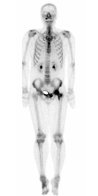
Fig. 3 Technetium-99m whole-body bone scintigraphy. Diffuse increased uptake throughout the left femoral head and intertrochanteric region is noted.
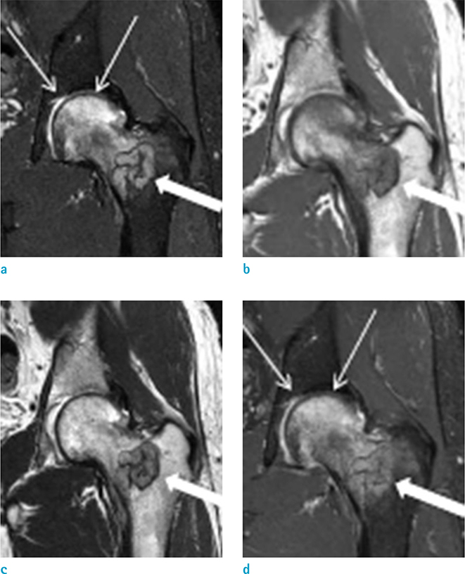
Fig. 4 Preoperative hip MRI. (a) Coronal fat-suppressed T2-weighted image displays a lobulating contoured mass (thick arrow) with a homogeneous nature and the absence of perilesional soft tissue or endosteal edema. Note the diffuse bone marrow edema, especially in the femoral head (thin arrows). (b) On the coronal T1-weighted image, the mass (thick arrow) exhibits an isointense signal intensity to the muscle. (c) On the coronal T2-weighted image, the mass exhibits a slight hyperintense relative to the skeletal muscle (thick arrow). (d) On the post-contrast fat-suppressed coronal T1-weighted image, the mass displays homogeneous enhancement (thick arrow) which indicates its solid nature. The adjacent endosteal bone and soft tissues are normal. No bone marrow edema or contrast enhancement in the femoral shaft adjacent or just inferior to the tumor was noted. The prominent marrow edema with mild contrast enhancement in the head portion of the femur (thin arrows) was believed to be due to the subchondral fracture, not the tumorous lesion itself.

Fig. 5 Microscopic examination of the local excised specimen. (a) Photomicrograph of a histologic specimen of schwannoma. Pathologic specimen (original magnification, × 400; Hematoxylin-Eosin stain) displays infiltrative growth of spindle cells forming abortive Verocay bodies (arrows) which have the characteristic findings of wavy, tightly organized, nuclear palisades with mild lymphocytic infiltration. (b) The brown staining was found in the cytoplasm, S-100 protein was positively expressed in the immunohistochemical staining (original magnification, × 400; S-100 stain).
Reference
-
1. Bansal AK, Bindal R, Shetty DC, Dua M. Rare occurrence of intraosseous schwannoma in a young child, its review and its pathogenesis. J Oral Maxillofac Pathol. 2012; 16:91–96.2. Simsek HO, Aksoy MC, Can C, Baykul T. Intraosseous schwannoma of the mandible. Int J Exper Dent Sci. 2012; 1:48–50.3. Sorensen SA, Mulvihill JJ, Nielsen A. Long-term follow-up of von Recklinghausen neurofibromatosis. Survival and malignant neoplasms. N Engl J Med. 1986; 314:1010–1015.4. Hoshi M, Takada J, Oebisu N, Nakamura H. Intraosseous schwannoma of the proximal femur. Asia Pac J Clin Oncol. 2012; 8:e29–e33.5. Gine J, Calmet J, Sirvent JJ, Domenech S. Intraosseous neurilemmoma of the radius: a case report. J Hand Surg Am. 2000; 25:365–369.6. Suzuki K, Yasuda T, Watanabe K, Kanamori M, Kimura T. Association between intraosseous schwannoma occurrence and the position of the intraosseous nutrient vessel: A case report. Oncol Lett. 2016; 11:3185–3188.7. Vora RA, Mintz DN, Athanasian EA. Intraosseous schwannoma of the metacarpal. Skeletal Radiol. 2000; 29:224–226.8. Imaizumi A, Kodama S, Sakamoto J, et al. Imaging findings of benign peripheral nerve sheath tumor in jaw. Oral Surg Oral Med Oral Pathol Oral Radiol. 2013; 116:369–376.9. Weiss SW, Goldblum JR, Folpe AL. Enzinger and Weiss's soft tissue tumors. Philadelphia, USA: Elsevier Health Sciences;2007.
