Associates and Prognosis of Giant Left Atrium; Single Center Experience
- Affiliations
-
- 1Division of Cardiology, Heart Center, Gangnam Severance Hospital, Yonsei University College of Medicine, Seoul, Korea. choi0928@yuhs.ac
- 2Division of Cardiology, Inje University College of Medicine, Busan, Korea.
- 3Division of Cardiology, Kyung Hee University School of Medicine, Seoul, Korea.
- KMID: 2392253
- DOI: http://doi.org/10.4250/jcu.2017.25.3.84
Abstract
- BACKGROUND
Left atrial (LA) remodeling develops as a result of longstanding pressure overload. However, determinants and clinical outcome of excessive remodeling, so called giant left atrium (GLA), are not clear.
METHODS
Clinical characteristics of patients with GLA (antero-posterior diameter higher than 65 mm), including echo-Doppler parameters, and follow-up clinical outcomes from a tertiary referral hospital were investigated.
RESULTS
Among 68519 consecutive primary patients who underwent echocardiography over a period of 10 years, data from 163 GLA cases (0.24%) were analyzed. Main causes were significant rheumatic mitral stenosis (n = 58, 36%); other causes comprised significant rheumatic mitral regurgitation (MR; n = 10, 6%), mitral valve (MV) prolapse or congenital mitral valvular disease (MVD) (n = 20, 12%), and functional MR (n = 25, 15%). However, mild rheumatic MV disease (n = 4, 3%) or left ventricular (LV) systolic or diastolic dysfunction without significant MR (n = 46, 28%) were also causes of GLA. During median follow-up of 22 months, 42 cases (26%) underwent composite events. MV surgery was related to lower rate of composite events. In multivariate analysis, MV surgery, elevated pulmonary arterial systolic pressure, and increased LA volume index were independent predictors of future events (p < 0.05) regardless of underlying diseases or history of MV surgery.
CONCLUSION
Although rheumatic MVD with atrial fibrillation is the main contributor to GLA, longstanding atrial fibrillation with LV dysfunction but without MVD also could be related to GLA. Even in GLA state, accurate measurement of LA volume is crucial for risk stratification for future events, regardless of underlying disease.
MeSH Terms
Figure
-
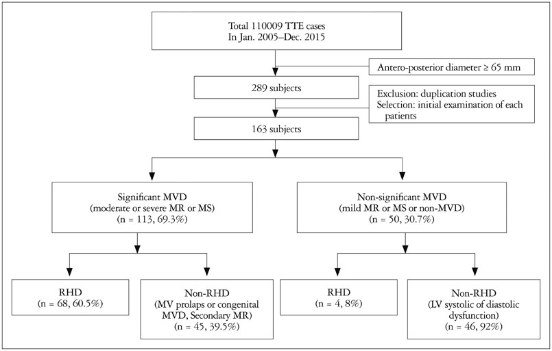
Fig. 1 Schematic flow diagram of the study population. TTE: transthoracic echocardiography, MR: mitral regurgitation, MS: mitral stenosis, MV: mitral valve, MVD: mitral valvular disease, RHD: rheumatic heart disease.
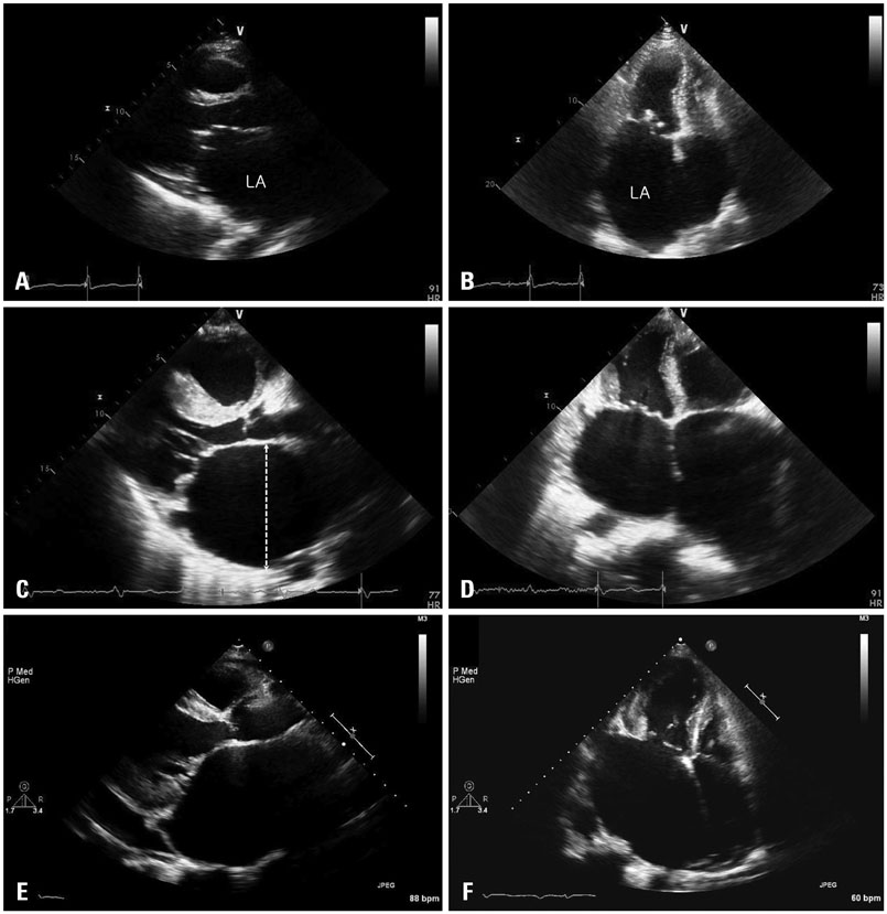
Fig. 2 Representative cases of a giant LA. Parasternal long axis view (A) and apical 4-chamber view (B) of rheumatic mitral stenosis. Parasternal long axis view (C) and apical 4-chamber view (D) of hypertrophic cardiomyopathy with moderate MR. Parasternal long axis view (E) and apical 4-chamber view (F) of severe rheumatic MR. Dotted line in C shows antero-posterior diameter of left atrium. LA: left atrium, MR: mitral regurgitation.
Cited by 1 articles
-
Cardiac Structural or Functional Changes Associated with CHA2DS2-VASc Scores in Nonvalvular Atrial Fibrillation: A Cross-Sectional Study Using Echocardiography
Albert Youngwoo Jang, Jongwook Yu, Ye Min Park, Mi Seung Shin, Wook-Jin Chung, Jeonggeun Moon
J Cardiovasc Imaging. 2018;26(3):135-143. doi: 10.4250/jcvi.2018.26.e17.
Reference
-
1. Abhayaratna WP, Seward JB, Appleton CP, Douglas PS, Oh JK, Tajik AJ, Tsang TS. Left atrial size: physiologic determinants and clinical applications. J Am Coll Cardiol. 2006; 47:2357–2363.2. De Jong AM, Maass AH, Oberdorf-Maass SU, Van Veldhuisen DJ, Van Gilst WH, Van Gelder IC. Mechanisms of atrial structural changes caused by stretch occurring before and during early atrial fibrillation. Cardiovasc Res. 2011; 89:754–765.3. Gerdts E, Wachtell K, Omvik P, Otterstad JE, Oikarinen L, Boman K, Dahlöf B, Devereux RB. Left atrial size and risk of major cardiovascular events during antihypertensive treatment: losartan intervention for end-point reduction in hypertension trial. Hypertension. 2007; 49:311–316.4. Manning WJ, Gelfand EV. Left atrial size and postoperative atrial fibrillation: the volume of evidence suggests it is time to break an old habit. J Am Coll Cardiol. 2006; 48:787–789.5. Laukkanen JA, Kurl S, Eränen J, Huttunen M, Salonen JT. Left atrium size and the risk of cardiovascular death in middle-aged men. Arch Intern Med. 2005; 165:1788–1793.6. El Maghraby A, Hajar R. Giant left atrium: a review. Heart Views. 2012; 13:46–52.7. Apostolakis E, Shuhaiber JH. The surgical management of giant left atrium. Eur J Cardiothorac Surg. 2008; 33:182–190.8. Oh JK. Echocardiographic evaluation of morphological and hemodynamic significance of giant left atrium. An important lesson. Circulation. 1992; 86:328–330.9. Lang RM, Badano LP, Mor-Avi V, Afilalo J, Armstrong A, Ernande L, Flachskampf FA, Foster E, Goldstein SA, Kuznetsova T, Lancellotti P, Muraru D, Picard MH, Rietzschel ER, Rudski L, Spencer KT, Tsang W, Voigt JU. Recommendations for cardiac chamber quantification by echocardiography in adults: an update from the American Society of Echocardiography and the European Association of Cardiovascular Imaging. Eur Heart J Cardiovasc Imaging. 2015; 16:233–270.10. Aurigemma GP, Gottdiener JS, Arnold AM, Chinali M, Hill JC, Kitzman D. Left atrial volume and geometry in healthy aging: the Cardiovascular Health Study. Circ Cardiovasc Imaging. 2009; 2:282–289.11. Jiamsripong P, Honda T, Reuss CS, Hurst RT, Chaliki HP, Grill DE, Schneck SL, Tyler R, Khandheria BK, Lester SJ. Three methods for evaluation of left atrial volume. Eur J Echocardiogr. 2008; 9:351–355.12. Grayburn PA, Weissman NJ, Zamorano JL. Quantitation of mitral regurgitation. Circulation. 2012; 126:2005–2017.13. Nishimura RA, Otto CM, Bonow RO, Carabello BA, Erwin JP 3rd, Guyton RA, O'Gara PT, Ruiz CE, Skubas NJ, Sorajja P, Sundt TM 3rd, Thomas JD. ACC/AHA Task Force Members. 2014 AHA/ACC Guideline for the Management of Patients With Valvular Heart Disease: a report of the American College of Cardiology/American Heart Association Task Force on Practice Guidelines. Circulation. 2014; 129:e521–e643.14. Plaschkes J, Borman JB, Merin G, Milwidsky H. Giant left atrium in rheumatic heart disease: a report of 18 cases treated by mitral valve replacement. Ann Surg. 1971; 174:194–201.15. Johnson J, Danielson GK, MacVaugh H 3rd, Joyner CR. Plication of the giant left atrium at operation for severe mitral regurgitation. Surgery. 1967; 61:118–121.16. Qian Y, Meng J, Tang H, Yang G, Deng Y, Wei D, Xiang B, Xiao X. Different structural remodelling in atrial fibrillation with different types of mitral valvular diseases. Europace. 2010; 12:371–377.17. Wang L, Di Tullio MR, Beecham A, Slifer S, Rundek T, Homma S, Blanton SH, Sacco RL. A comprehensive genetic study on left atrium size in Caribbean Hispanics identifies potential candidate genes in 17p10. Circ Cardiovasc Genet. 2010; 3:386–392.18. Matsuda H, Nakao M, Nohara H, Higami T, Mukohara N, Asada T, Ogawa K, Kawamura T. [The causes of prolonged postoperative respiratory care in mitral valve disease with a giant left atrium]. Kyobu Geka. 1990; 43:172–177.19. Kawazoe K, Beppu S, Takahara Y, Nakajima N, Tanaka K, Ichihashi K, Fujita T, Manabe H. Surgical treatment of giant left atrium combined with mitral valvular disease. Plication procedure for reduction of compression to the left ventricle, bronchus, and pulmonary parenchyma. J Thorac Cardiovasc Surg. 1983; 85:885–892.20. Tsang TS, Abhayaratna WP, Barnes ME, Miyasaka Y, Gersh BJ, Bailey KR, Cha SS, Seward JB. Prediction of cardiovascular outcomes with left atrial size: is volume superior to area or diameter? J Am Coll Cardiol. 2006; 47:1018–1023.21. Mahabadi AA, Samy B, Seneviratne SK, Toepker MH, Bamberg F, Hoffmann U, Truong QA. Quantitative assessment of left atrial volume by electrocardiographic-gated contrast-enhanced multidetector computed tomography. J Cardiovasc Comput Tomogr. 2009; 3:80–87.
- Full Text Links
-
- Actions
-
Cited
- CITED
-
- Close
- Share
-
- Similar articles
-
- A Giant Left Atrium in Rheumatic Mitral Stenosis
- A Case of Giant Condyloma of the Penis
- Giant Aneurysm of a Congenital Coronary Arteriovenous Fistula Arising from the Left Coronary Artery
- A Case of Recurrent Thrombus Associated with Left Atrial Calcification
- A Case of Primary Leiomyosarcoma with Prominent Osteoclast-like Giant Cell of Lung with Cardiac Invasion
