Finite element analysis of maxillary incisor displacement during en-masse retraction according to orthodontic mini-implant position
- Affiliations
-
- 1Department of Orthodontics, Graduate School of Clinical Dental Science, The Catholic University of Korea, Seoul, Korea.
- 2Private Practice, Seoul, Korea.
- 3Department of Orthodontics, College of Dentistry, Yonsei University, Seoul, Korea.
- 4Division of Orthodontics, Department of Dentistry, Asan Medical Center, Seoul, Korea.
- 5Department of Orthodontics, Ewha Womans University Mokdong Hospital, Seoul, Korea.
- 6Division of Orthodontics, Department of Dentistry, St. Paul's Hospital, College of Medicine, The Catholic University of Korea, Seoul, Korea. dmoss1@hanmail.net
- KMID: 2392204
- DOI: http://doi.org/10.4041/kjod.2016.46.4.242
Abstract
OBJECTIVE
Orthodontic mini-implants (OMI) generate various horizontal and vertical force vectors and moments according to their insertion positions. This study aimed to help select ideal biomechanics during maxillary incisor retraction by varying the length in the anterior retraction hook (ARH) and OMI position.
METHODS
Two extraction models were constructed to analyze the three-dimentional finite element: a first premolar extraction model (Model 1, M1) and a residual 1-mm space post-extraction model (Model 2, M2). The OMI position was set at a height of 8 mm from the arch wire between the second maxillary premolar and the first molar (low OMI traction) or at a 12-mm height in the mesial second maxillary premolar (high OMI traction). Retraction force vectors of 200 g from the ARH (-1, +1, +3, and +6 mm) at low or high OMI traction were resolved into X-, Y-, and Z-axis components.
RESULTS
In M1 (low and high OMI traction) and M2 (low OMI traction), the maxillary incisor tip was extruded, but the apex was intruded, and the occlusal plane was rotated clockwise. Significant intrusion and counter-clockwise rotation in the occlusal plane were observed under high OMI traction and -1 mm ARH in M2.
CONCLUSIONS
This study observed orthodontic tooth movement according to the OMI position and ARH height, and M2 under high OMI traction with short ARH showed retraction with maxillary incisor intrusion.
Keyword
Figure
-
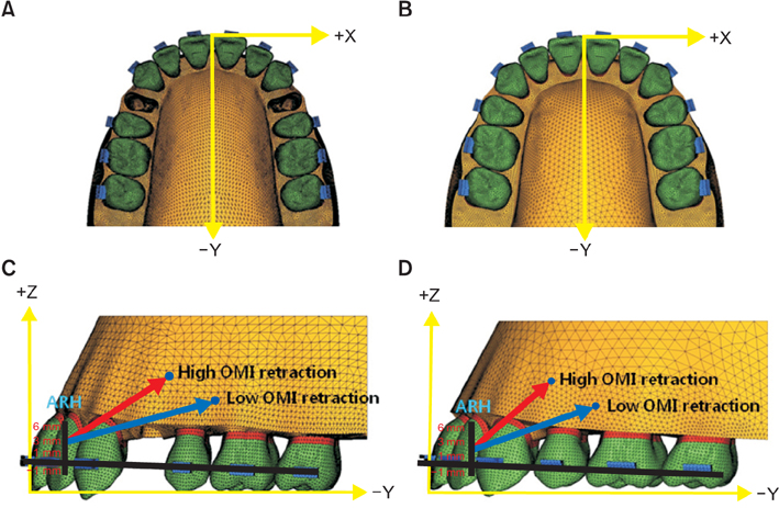
Figure 1 Three-dimensional finite element models. A, Occlusal view of the model with extraction of the first premolar (Model 1, M1). B, Occlusal view of the model with residual extraction space of 1 mm (Model 2, M2). C, Lateral view of the model with extraction of the first premolar (M1). D, Lateral view of the model with residual extraction space of 1 mm (M2). ARH, Anterior retraction hook; OMI, orthodontic mini-implants; X, medio-lateral; +, lateral; -, medial direction; Y, anterio-posterior; +, anterior; -, posterior direction; Z, superio-inferior; +, superior; -, inferior direction.
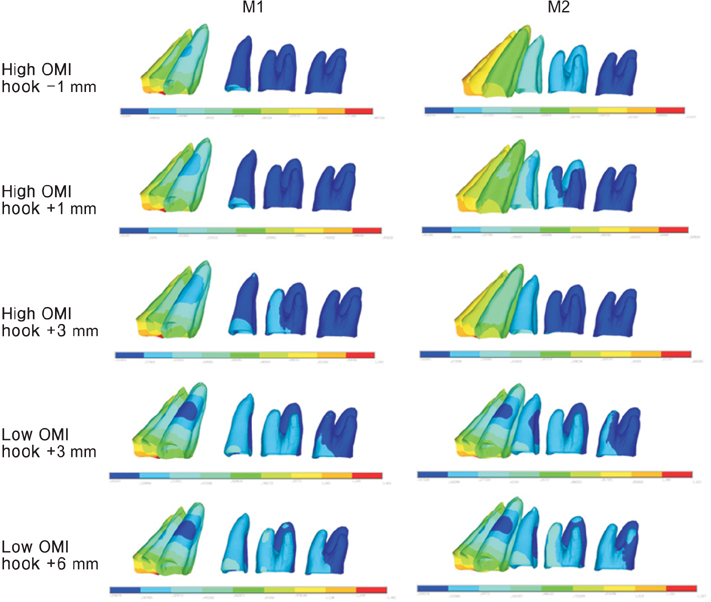
Figure 2 Von Mises stress distribution (g/mm2). M1, Model 1; M2, Model 2; OMI, orthodontic mini-implants.

Figure 3 The axis changes of the maxillary central incisors according to the length of the anterior retraction hook under low and high orthodontic mini-implant traction in both models with extraction of the first premolar (Model 1, M1) and a residual extraction space of 1 mm (Model 2, M2). Solid line, original axis of the maxillary central incisor; dashed line, changed axis of the maxillary central incisor; ○, -1 mm hook; □, +1 mm hook; △, + 3 mm hook; ×, +6 mm hook.

Figure 4 The axis change in the maxillary central incisor retracted by low and high orthodontic mini-implant (OMI) traction with a 3-mm anterior retraction hook in both models with extraction of the first premolar (Model 1, M1) and a residual extraction space of 1 mm (Model 2, M2). Solid line, original axis of the maxillary central incisor; dashed line, changed axis of the maxillary central incisor; ○, low OMI retraction; □, high OMI retraction.
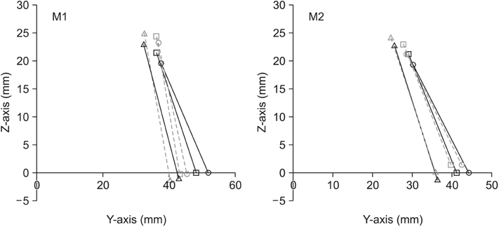
Figure 5 The axis change in the maxillary incisors retracted by high orthodontic mini-implant traction with a -1 mm anterior retraction hook in both models with extraction of the first premolar (Model 1, M1) and a residual extraction space of 1 mm (Model 2, M2). Solid line, original axis of maxillary incisors; dashed line, changed axis of maxillary incisors; ○, maxillary central incisor; □, maxillary lateral incisor; △, maxillary canine.
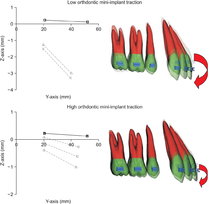
Figure 6 Movement pattern of the model with extraction of the first premolar (Model 1, M1). The rotation of the occlusal plane according to the length of anterior retraction hook used in the model with extraction of the first premolar (M1) under low and high orthodontic miniimplant traction. Solid line, original occlusal plane; dashed line, rotated occlusal plane; ○, -1 mm hook; □, +1 mm hook; △, +3 mm hook; ×, +6 mm hook.
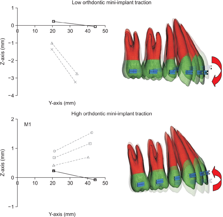
Figure 7 Movement pattern of the model with residual extraction space of 1 mm (Model 2, M2). The rotation of the occlusal plane according to the length of the anterior retraction hook used in the model with residual extraction space of 1 mm (M2) under low and high orthodontic mini-implant traction. Solid line, original occlusal plane; dashed line, rotated occlusal plane; ○, -1 mm hook; □, +1 mm hook; △, +3 mm hook; ×, +6 mm hook.
Cited by 2 articles
-
Evaluation of the effects of miniscrew incorporation in palatal expanders for young adults using finite element analysis
Eui-Hyang Seong, Sung-Hwan Choi, Hee-Jin Kim, Hyung-Seog Yu, Young-Chel Park, Kee-Joon Lee
Korean J Orthod. 2018;48(2):81-89. doi: 10.4041/kjod.2018.48.2.81.Effect of archwire stiffness and friction on maxillary posterior segment displacement during anterior segment retraction: A three-dimensional finite element analysis
Choon-Soo Park, Hyung-Seog Yu, Jung-Yul Cha, Sung-Seo Mo, Kee-Joon Lee
Korean J Orthod. 2019;49(6):393-403. doi: 10.4041/kjod.2019.49.6.393.
Reference
-
1. Cha BK, Lee JY, Jost-Brinkmann PG, Yoshida N. Analysis of tooth movement in extraction cases using three-dimensional reverse engineering technology. Eur J Orthod. 2007; 29:325–331.
Article2. Cho MY, Choi JH, Lee SP, Baek SH. Three-dimensional analysis of the tooth movement and arch dimension changes in Class I malocclusions treated with first premolar extractions: a guideline for virtual treatment planning. Am J Orthod Dentofacial Orthop. 2010; 138:747–757.
Article3. Lai EH, Yao CC, Chang JZ, Chen I, Chen YJ. Three-dimensional dental model analysis of treatment outcomes for protrusive maxillary dentition: comparison of headgear, miniscrew, and miniplate skeletal anchorage. Am J Orthod Dentofacial Orthop. 2008; 134:636–645.
Article4. Upadhyay M, Yadav S, Nagaraj K, Patil S. Treatment effects of mini-implants for en-masse retraction of anterior teeth in bialveolar dental protrusion patients: a randomized controlled trial. Am J Orthod Dentofacial Orthop. 2008; 134:18–29.e1.
Article5. Upadhyay M, Yadav S, Patil S. Mini-implant anchorage for en-masse retraction of maxillary anterior teeth: a clinical cephalometric study. Am J Orthod Dentofacial Orthop. 2008; 134:803–810.
Article6. Lim JK. Gummy smile correction using mini implant. Korean J Clin Orthod. 2010; 9:14–31.7. Lee KJ, Park YC, Hwang CJ, Kim YJ, Choi TH, Yoo HM, et al. Displacement pattern of the maxillary arch depending on miniscrew position in sliding mechanics. Am J Orthod Dentofacial Orthop. 2011; 140:224–232.
Article8. Kim SJ, Chun YS, Jung SH, Park SH. Three dimensional analysis of tooth movement using different types of maxillary molar distalization appliances. Korean J Orthod. 2008; 38:376–387.
Article9. Lee HA, Park YC. Treatment and posttreatment changes following intrusion of maxillary posterior teeth with miniscrew implants for open bite correction. Korean J Orthod. 2008; 38:31–40.
Article10. Mo SS, Kim SH, Sung SJ, Chung KR, Chun YS, Kook YA, et al. Factors controlling anterior torque with C-implants depend on en-masse retraction without posterior appliances: biocreative therapy type II technique. Am J Orthod Dentofacial Orthop. 2011; 139:e183–e191.
Article11. Tominaga JY, Tanaka M, Koga Y, Gonzales C, Kobayashi M, Yoshida N. Optimal loading conditions for controlled movement of anterior teeth in sliding mechanics. Angle Orthod. 2009; 79:1102–1107.
Article12. Sia S, Shibazaki T, Koga Y, Yoshida N. Experimental determination of optimal force system required for control of anterior tooth movement in sliding mechanics. Am J Orthod Dentofacial Orthop. 2009; 135:36–41.
Article13. Kojima Y, Fukui H. A finite element simulation of initial movement, orthodontic movement, and the centre of resistance of the maxillary teeth connected with an archwire. Eur J Orthod. 2014; 36:255–261.
Article14. Chong DR, Jang YJ, Chun YS, Jung SH, Lee SK. The evaluation of rotational movements of maxillary posterior teeth using three dimensional images in cases of extraction of maxillary first premolar. Korean J Orthod. 2005; 35:451–458.15. Jeong GM, Sung SJ, Lee KJ, Chun YS, Mo SS. Finite-element investigation of the center of resistance of the maxillary dentition. Korean J Orthod. 2009; 39:83–94.
Article16. Germane N, Bentley BE Jr, Isaacson RJ. Three biologic variables modifying faciolingual tooth angulation by straight-wire appliances. Am J Orthod Dentofacial Orthop. 1989; 96:312–319.
Article17. Andrews LF. The straight-wire appliance. Explained and compared. J Clin Orthod. 1976; 10:174–195.18. Coolidge ED. The thickness of the human periodontal membrane. J Am Dent Assoc Dent Cosm. 1937; 24:1260–1270.
Article19. Tanne K, Sakuda M, Burstone CJ. Three-dimensional finite element analysis for stress in the periodontal tissue by orthodontic forces. Am J Orthod Dentofacial Orthop. 1987; 92:499–505.
Article20. Sung EH, Kim SJ, Chun YS, Park YC, Yu HS, Lee KJ. Distalization pattern of whole maxillary dentition according to force application points. Korean J Orthod. 2015; 45:20–28.
Article21. Marcuschamer E, Tsukiyama T, Griffin TJ, Arguello E, Gallucci GO, Magne P. Anatomical crown width/length ratios of worn and unworn maxillary teeth in Asian subjects. Int J Periodontics Restorative Dent. 2011; 31:495–503.22. Brook AH, Holt RD. The relationship of crown length to root length in permanent maxillary central incisors. Proc Br Paedod Soc. 1978; 8:17–20.23. Sia S, Koga Y, Yoshida N. Determining the center of resistance of maxillary anterior teeth subjected to retraction forces in sliding mechanics. An in vivo study. Angle Orthod. 2007; 77:999–1003.
Article24. Sung SJ, Jang GW, Chun YS, Moon YS. Effective en-masse retraction design with orthodontic mini-implant anchorage: a finite element analysis. Am J Orthod Dentofacial Orthop. 2010; 137:648–657.
Article25. van Steenbergen E, Burstone CJ, Prahl-Andersen B, Aartman IH. The relation between the point of force application and flaring of the anterior segment. Angle Orthod. 2005; 75:730–735.26. Choy K, Kim KH, Burstone CJ. Initial changes of centres of rotation of the anterior segment in response to horizontal forces. Eur J Orthod. 2006; 28:471–474.
Article27. Park HS, Lee SK, Kwon OW. Group distal movement of teeth using microscrew implant anchorage. Angle Orthod. 2005; 75:602–609.28. Park HM, Kim BH, Yang IH, Baek SH. Preliminary three-dimensional analysis of tooth movement and arch dimension change of the maxillary dentition in Class II division 1 malocclusion treated with first premolar extraction: conventional anchorage vs. mini-implant anchorage. Korean J Orthod. 2012; 42:280–290.
Article
- Full Text Links
-
- Actions
-
Cited
- CITED
-
- Close
- Share
-
- Similar articles
-
- Factors influencing the axes of anterior teeth during SWA en masse sliding retraction with orthodontic mini-implant anchorage: a finite element study
- Three dimensional finite element analysis of the phenomenon during distal en masse movement of the maxillary dentition
- The pattern of movement and stress distribution during retraction of maxillary incisors using a 3-D finite element method
- Palatal en-masse retraction of segmented maxillary anterior teeth: A finite element study
- A study on the pattern of movement during retraction of maxillary central incisor by finite element method
