Comparisons of orthodontic root resorption under heavy and jiggling reciprocating forces during experimental tooth movement in a rat model
- Affiliations
-
- 1Department of Orthodontics, Nihon University School of Dentistry at Matsudo, Japan. yamaguchi.masaru@nihon-u.ac.jp
- KMID: 2392203
- DOI: http://doi.org/10.4041/kjod.2016.46.4.228
Abstract
OBJECTIVE
Root mobility due to reciprocating movement of the tooth (jiggling) may exacerbate orthodontic root resorption (ORR). "Jiggling" describes mesiodistal or buccolingual movement of the roots of the teeth during orthodontic treatment. In the present study, buccolingual movement is described as "jiggling." We aimed to investigate the relationship between ORR and jiggling and to test for positive cell expression in odontoclasts in resorbed roots during experimental tooth movement (jiggling) in vivo.
METHODS
Male Wistar rats were divided into control, heavy force (HF), optimal force (OF), and jiggling force (JF) groups. The expression levels of cathepsin K, matrix metalloproteinase (MMP)-9 protein, interleukin (IL)-6, cytokine-induced neutrophil chemoattractant 1 (CINC-1; an IL-8-related protein in rodents), receptor activator of nuclear factor κB ligand (RANKL), and osteoprotegerin protein in the dental root were determined using immunohistochemistry.
RESULTS
On day 21, a greater number of root resorption lacunae, which contained multinucleated odontoclasts, were observed in the palatal roots of rats in the JF group than in rats from other groups. Furthermore, there was a significant increase in the numbers of cathepsin K-positive and MMP-9-positive odontoclasts in the JF group on day 21. Immunoreactivities for IL-6, CINC-1, and RANKL were stronger in resorbed roots exposed to jiggling than in the other groups on day 21. Negative reactivity was observed in the controls.
CONCLUSIONS
These results suggest that jiggling may induce ORR via inflammatory cytokine production during orthodontic tooth movement, and that jiggling may be a risk factor for ORR.
MeSH Terms
Figure
-

Figure 1 Experimental tooth movement. Experimental tooth movement was induced by the design of an appliance (diameter: 0.012 inches [0.3048 mm], stainless steel wire) ligated to a maxillary first molar cleat by a 0.008-inches (0.2032 mm) stainless steel ligature wire. The upper first molar was moved palatally or buccally using the appliance with a force of 10 g or 50 g. The appliances were attached to the rats after activation in each direction.
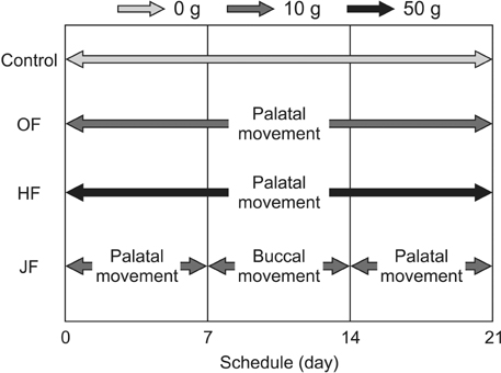
Figure 2 The experimental schedule for each group. The rats were randomly assigned to four groups: the control group with no appliance (days 0–21); the optimal force (OF) group, treated with 10 g of compression (days 0–21); the heavy force (HF) group, treated with 50 g of compression (days 0–21); and the jiggling force (JF) group, treated with 10 g compression on day 7 (days 0–7), 10 g of tension on day 14 (days 8–14), and 10 g of compression (the A area) on day 21 (days 15–21); and 10 g of tension on day 7 (days 0–7), 10 g of compression on day 14 (days 8–14), and 10 g of tension (the B area) on day 21 (days 15–21).

Figure 3 A schematic illustration showing the area of investigation (box) on the palatal aspect of the distal palatal root of the first molar (hematoxylin and eosin staining). MP, Mesial palatal root; M, mesial root; MB, mesial buccal root; DP, distal palatal root; DB, distal buccal root; M1, the left first upper molar; PDL, periodontal ligament.

Figure 4 Light microscopy images showing the effects of different orthodontic forces on multinucleated osteoclasts (hematoxylin and eosin staining, ×400). The expression of odontoclasts (arrowheads) on the cementum in the jiggling force group (u) was greater than those in the heavy force (o) and optimal force groups (i) on day 21. The direction of the applied force is indicated by the large arrow. PDL, Periodontal ligament; C, cementum; D, dentin.

Figure 5 Effects of different orthodontic forces on the expression of cathepsin K-positive odontoclasts (×400). PDL, Periodontal ligament; C, cementum; D, dentin.

Figure 6 Effects of different orthodontic forces on the expression of matrix metalloproteinase (MMP)-9-positive odontoclasts (×400). PDL, Periodontal ligament; C, cementum; D, dentin.

Figure 7 Effects of different orthodontic forces on the expression of interleukin (IL)-6-positive odontoclasts (×400). PDL, Periodontal ligament; C, cementum; D, dentin.

Figure 8 Effects of different orthodontic forces on the expression of cytokine-induced neutrophil chemoattractant 1 (CINC-1)-positive odontoclasts (×400). PDL, Periodontal ligament; C, cementum; D, dentin.

Figure 9 Effects of different orthodontic forces on the expression of receptor activator of nuclear factor κB ligand (RANKL)-positive odontoclasts (×400). PDL, Periodontal ligament; C, cementum; D, dentin.

Figure 10 Effects of different orthodontic forces on the expression of osteoprotegerin (OPG)-positive odontoclasts (×400). PDL, Periodontal ligament; C, cementum; D, dentin.

Figure 11 A statistical diagram depicting the quantitative assessment of cellular changes. MMP, Matrix metalloproteinase; IL, interleukin; CINC-1, cytokine-induced neutrophil chemoattractant 1; RANKL, receptor activator of nuclear factor κB ligand; OPG, osteoprotegerin; Control, control group; OF, optimal force group; HF, heavy force group; JF, jiggling force group. *p < 0.05, **p < 0.01, significantly different from the corresponding control group; †p < 0.05, ††p < 0.01, significantly different from the corresponding HF.
Cited by 2 articles
-
Cone-beam computed tomography for the assessment of root–crown ratios of the maxillary and mandibular incisors in a Korean population
Sung-Hwan Choi, Jung-Suk Kim, Cheol-Soon Kim, Hyung-Seog Yu, Chung-Ju Hwang
Korean J Orthod. 2017;47(1):39-49. doi: 10.4041/kjod.2017.47.1.39.Effect of caspases and RANKL induced by heavy force in orthodontic root resorption
Yukari Minato, Masaru Yamaguchi, Mami Shimizu, Jun Kikuta, Takuji Hikida, Momoko Hikida, Masaaki Suemitsu, Kayo Kuyama, Kazutaka Kasai
Korean J Orthod. 2018;48(4):253-261. doi: 10.4041/kjod.2018.48.4.253.
Reference
-
1. Kaley J, Phillips C. Factors related to root resorption in edgewise practice. Angle Orthod. 1991; 61:125–132.2. Malmgren O, Goldson L, Hill C, Orwin A, Petrini L, Lundberg M. Root resorption after orthodontic treatment of traumatized teeth. Am J Orthod. 1982; 82:487–491.
Article3. Linge BO, Linge L. Apical root resorption in upper anterior teeth. Eur J Orthod. 1983; 5:173–183.
Article4. Owman-Moll P, Kurol J, Lundgren D. The effects of a four-fold increased orthodontic force magnitude on tooth movement and root resorptions. An intraindividual study in adolescents. Eur J Orthod. 1996; 18:287–294.
Article5. Owman-Moll P, Kurol J, Lundgren D. Effects of a doubled orthodontic force magnitude on tooth movement and root resorptions. An inter-individual study in adolescents. Eur J Orthod. 1996; 18:141–150.
Article6. Chan E, Darendeliler MA. Physical properties of root cementum: Part 5. Volumetric analysis of root resorption craters after application of light and heavy orthodontic forces. Am J Orthod Dentofacial Orthop. 2005; 127:186–195.
Article7. Brezniak N, Wasserstein A. Root resorption after orthodontic treatment: Part 2. Literature review. Am J Orthod Dentofacial Orthop. 1993; 103:138–146.
Article8. Mirabella AD, Artun J. Risk factors for apical root resorption of maxillary anterior teeth in adult orthodontic patients. Am J Orthod Dentofacial Orthop. 1995; 108:48–55.
Article9. Hayashi A, Hayashi H, Kawata T. Prevention of root resorption in hypofunctional teeth by occlusal function recovery. Angle Orthod. 2015; DOI: 10.2319/012215-47.1. [Epub ahead of print].
Article10. Nakano Y, Yamaguchi M, Fujita S, Asano M, Saito K, Kasai K. Expressions of RANKL/RANK and M-CSF/c-fms in root resorption lacunae in rat molar by heavy orthodontic force. Eur J Orthod. 2011; 33:335–343.
Article11. Kaku M, Sumi H, Shikata H, Kojima S, Motokawa M, Fujita T, et al. Effects of pulpectomy on the amount of root resorption during orthodontic tooth movement. J Endod. 2014; 40:372–378.
Article12. Gonzales C, Hotokezaka H, Yoshimatsu M, Yozgatian JH, Darendeliler MA, Yoshida N. Force magnitude and duration effects on amount of tooth movement and root resorption in the rat molar. Angle Orthod. 2008; 78:502–509.
Article13. Kohno T, Matsumoto Y, Kanno Z, Warita H, Soma K. Experimental tooth movement under light orthodontic forces: rates of tooth movement and changes of the periodontium. J Orthod. 2002; 29:129–135.
Article14. Gameiro GH, Nouer DF, Pereira-Neto JS, de Araújo Magnani MB, de Andrade ED, Novaes PD, et al. Histological analysis of orthodontic root resorption in rats treated with the cyclooxygenase-2 (COX-2) inhibitor celecoxib. Orthod Craniofac Res. 2008; 11:156–161.
Article15. Eross E, Turk T, Elekdag-Turk S, Cakmak F, Jones AS, Végh A, et al. Physical properties of root cementum: Part 25. Extent of root resorption after the application of light and heavy buccopalatal jiggling forces for 12 weeks: A microcomputed tomography study. Am J Orthod Dentofacial Orthop. 2015; 147:738–746.
Article16. Brudvik P, Rygh P. Non-clast cells start orthodontic root resorption in the periphery of hyalinized zones. Eur J Orthod. 1993; 15:467–480.
Article17. Brezniak N, Wasserstein A. Root resorption after orthodontic treatment: Part 1. Literature review. Am J Orthod Dentofacial Orthop. 1993; 103:62–66.
Article18. Brudvik P, Rygh P. Root resorption beneath the main hyalinized zone. Eur J Orthod. 1994; 16:249–263.
Article19. Kurol J, Owman-Moll P. Hyalinization and root resorption during early orthodontic tooth movement in adolescents. Angle Orthod. 1998; 68:161–165.20. Kvam E. Scanning electron microscopy of tissue changes on the pressure surface of human premolars following tooth movement. Scand J Dent Res. 1972; 80:357–368.
Article21. Rygh P. Ultrastructural cellular reactions in pressure zones of rat molar periodontium incident to orthodontic tooth movement. Acta Odontol Scand. 1972; 30:575–593.
Article22. Ohashi M, Yamaguchi M, Hikida T, Kikuta J, Shimizu M, Goseki T, et al. Jiggling force induces orthodontic root resorption during tooth movement in rats. Int Oral-Med Sci. 2015; 14:13–20.
Article23. Meikle MC. The tissue, cellular, and molecular regulation of orthodontic tooth movement: 100 years after Carl Sandstedt. Eur J Orthod. 2006; 28:221–240.
Article24. Wucherpfennig AL, Li YP, Stetler-Stevenson WG, Rosenberg AE, Stashenko P. Expression of 92 kD type IV collagenase/gelatinase B in human osteoclasts. J Bone Miner Res. 1994; 9:549–556.
Article25. Oshiro T, Shibasaki Y, Martin TJ, Sasaki T. Immunolocalization of vacuolar-type H+-ATPase, cathepsin K, matrix metalloproteinase-9, and receptor activator of NFkappaB ligand in odontoclasts during physiological root resorption of human deciduous teeth. Anat Rec. 2001; 264:305–311.
Article26. Tsuchiya M, Akiba Y, Takahashi I, Sasano Y, Kashiwazaki J, Tsuchiya S, et al. Comparison of expression patterns of cathepsin K and MMP-9 in odontoclasts and osteoclasts in physiological root resorption in the rat molar. Arch Histol Cytol. 2008; 71:89–100.
Article27. Asano M, Yamaguchi M, Nakajima R, Fujita S, Utsunomiya T, Yamamoto H, et al. IL-8 and MCP-1 induced by excessive orthodontic force mediates odontoclastogenesis in periodontal tissues. Oral Dis. 2011; 17:489–498.
Article28. Hayashi N, Yamaguchi M, Nakajima R, Utsunomiya T, Yamamoto H, Kasai K. T-helper 17 cells mediate the osteo/odontoclastogenesis induced by excessive orthodontic forces. Oral Dis. 2012; 18:375–388.
Article29. Kikuta J, Yamaguchi M, Shimizu M, Yoshino T, Kasai K. Notch signaling induces root resorption via RANKL and IL-6 from hPDL cells. J Dent Res. 2015; 94:140–147.
Article30. Garlet TP, Coelho U, Repeke CE, Silva JS, Cunha Fde Q, Garlet GP. Differential expression of osteoblast and osteoclast chemmoatractants in compression and tension sides during orthodontic movement. Cytokine. 2008; 42:330–335.
Article
- Full Text Links
-
- Actions
-
Cited
- CITED
-
- Close
- Share
-
- Similar articles
-
- Changes in the titer of tooth root antibodies accompanying root resorption associated with orthodontic tooth movement
- Root resorption and bone resorption by jiggling force in cat premolars
- A study of root resorption and alveolar bone changes during tooth movement after treatment with etidronate disodium
- Mechanisms of Osteoclastogenesis in Orthodontic Tooth Movement and Orthodontically Induced Tooth Root Resorption
- A study on the affecting factors on root resorption
