Total intrusion and distalization of the maxillary arch to improve smile esthetics
- Affiliations
-
- 1Department of Orthodontics, Gangnam Severance Hospital, College of Dentistry, Yonsei University, Seoul, Korea. crchung@yuhs.ac
- 2The Institute of Craniofacial Deformity, College of Dentistry, Yonsei University, Seoul, Korea.
- KMID: 2392197
- DOI: http://doi.org/10.4041/kjod.2017.47.1.59
Abstract
- This case report illustrates the successful treatment of a patient with skeletal Class II malocclusion and an unesthetic smile involving excessive gingival display and large buccal corridors. By applying dual buccal interradicular miniscrews, total intrusion of the maxillary dentition along with distalization was induced to improve both the occlusion and smile esthetics. In addition to the conventional cephalometric superimposition, three-dimensional superimposition was performed and evaluated to validate the treatment outcome.
Figure
-
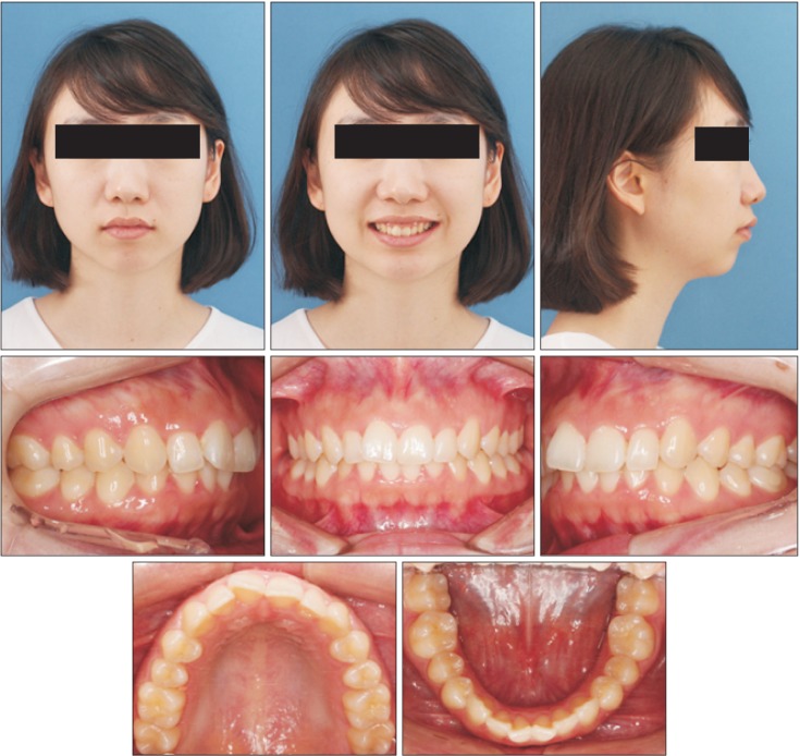
Figure 1 Pretreatment facial and intraoral photographs.
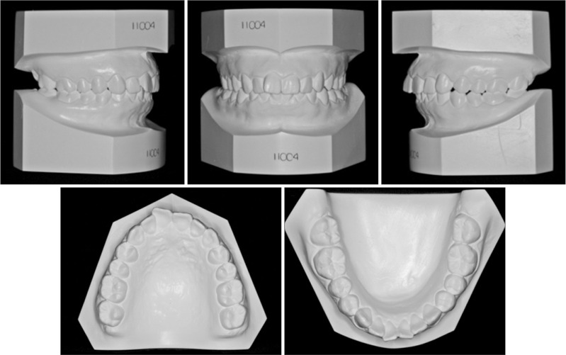
Figure 2 Pretreatment dental casts.
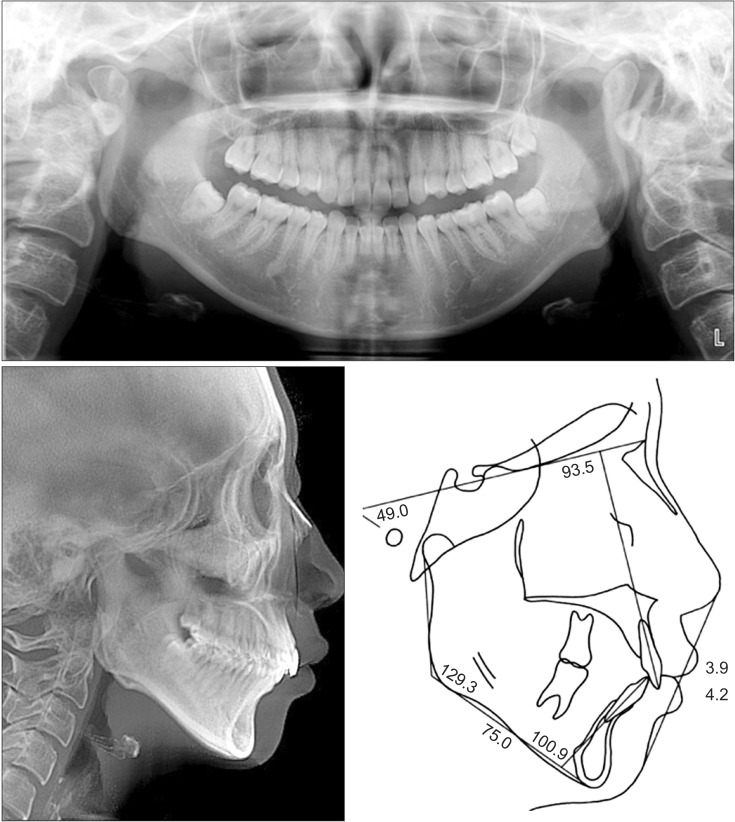
Figure 3 Pretreatment radiographs and cephalometric tracing.
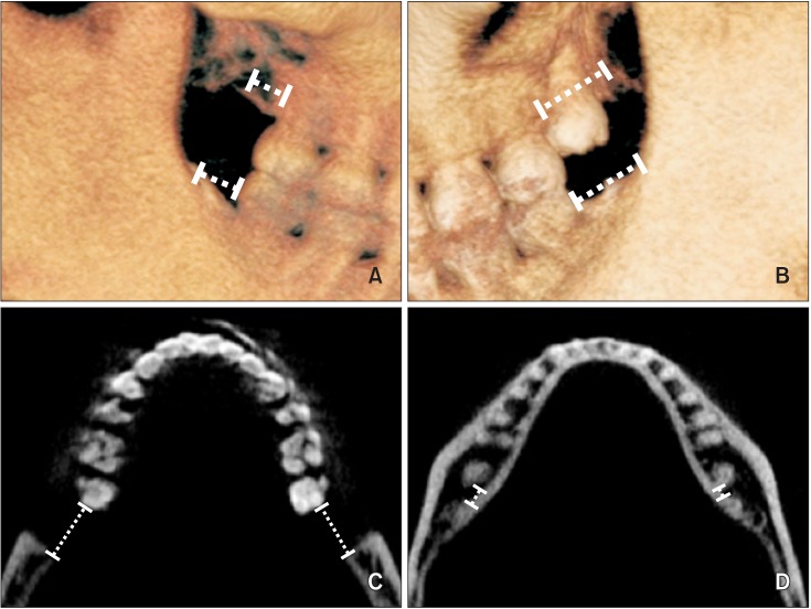
Figure 4 Posterior anatomic limit for total distalization. A and B, The available space at the maxillary tuberosity distal to the second molars, and the available space between the mandibular second molar and the anterior border of the ramus in the sagittal plane; C and D, the available space between the second molar crown to the anterior border of the ramus and between the root to the inner lingual cortex of the mandible in the axial plane.
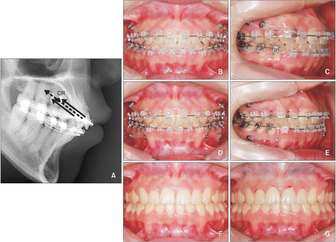
Figure 5 Biomechanical diagram and intraoral photographs. A, Line of force passing through the center of resistance (CR); B and C, dual miniscrews used for total intrusion and distalization of the maxillary arch; D and E, additional miniscrews inserted in the mandible for total distalization of the mandibular arch. F and G, Intraoral photographs before and after gingivoplasty.
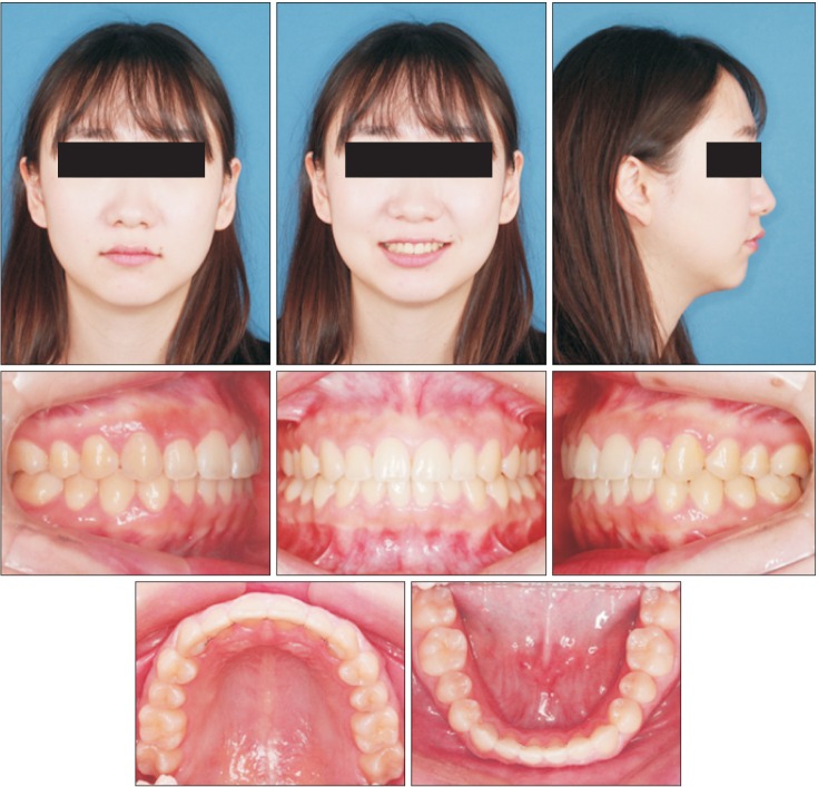
Figure 6 Posttreatment facial and intraoral photographs.
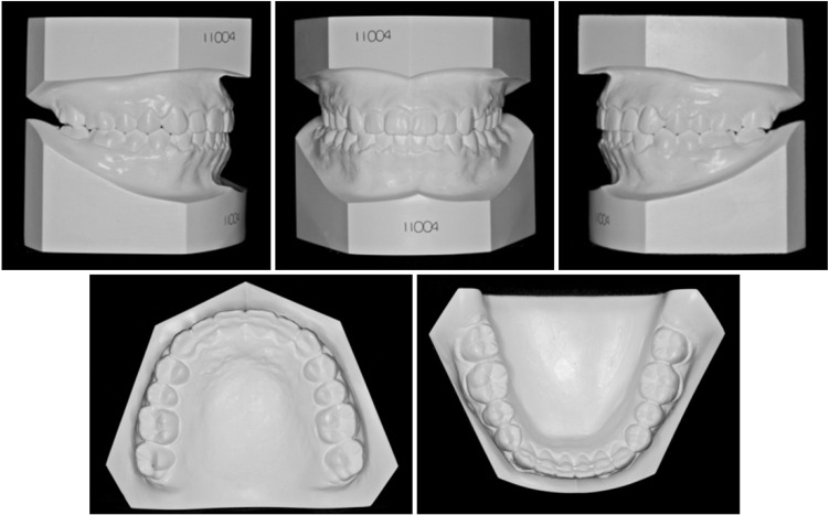
Figure 7 Posttreatment dental casts.
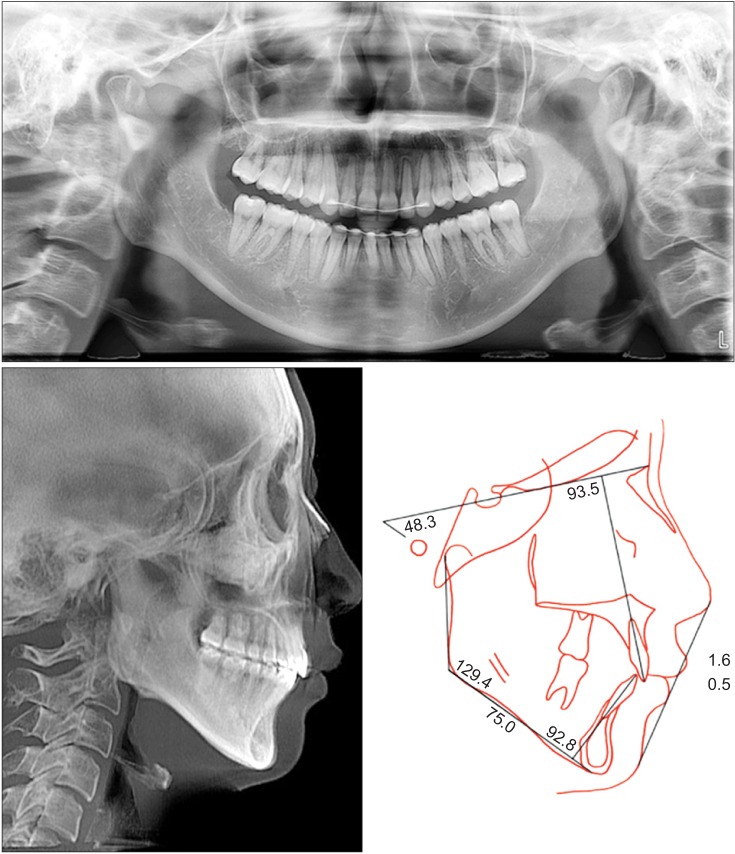
Figure 8 Posttreatment radiographs and cephalometric tracing.
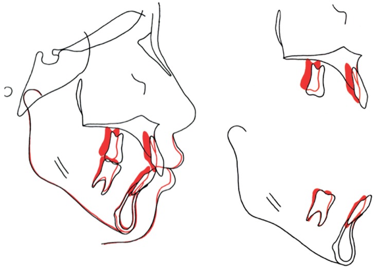
Figure 9 Cephalometric superimposition. Black, initial; red, final.
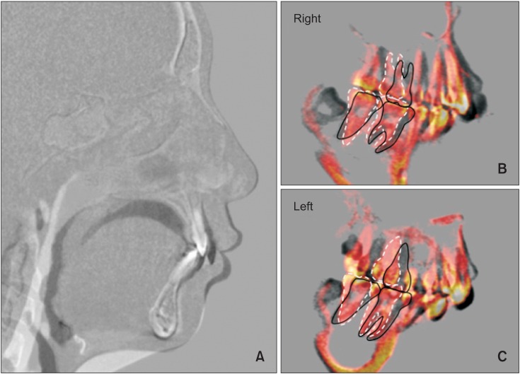
Figure 10 Three-dimensional cone-beam computed tomography superimposition to the cranial base. A, Sagittal cut near the midsagittal plane showing the mesial margins of the maxillary right central incisors. Incisal intrusion and distalization along with the changes in the lips are noted. Black, initial; white, final. B and C, Sagittal cut along the posterior arch form connecting the midroot portion of each tooth showing the movement of the maxillary and mandibular posterior teeth. Translation of the maxillary molars and the distal tip of the mandibular molars are noted. Black solid line, initial; white dotted line, final.
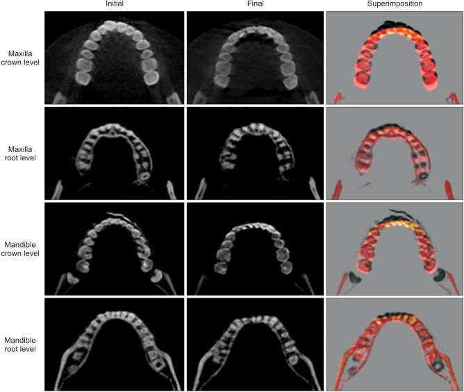
Figure 11 Changes in the arch form and tooth position following treatment. Axial cuts were made at the mid-crown level and midroot level before and after treatment. Black, initial; white, final.
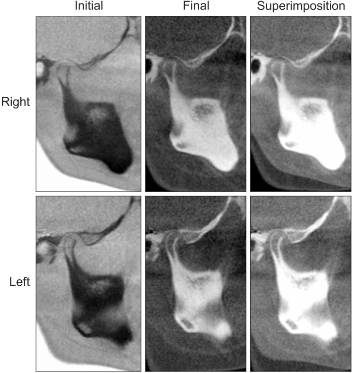
Figure 12 Three-dimensional cone-beam computed tomography superimposition of the temporomandibular joint. No distinct changes in condylar position are noted.
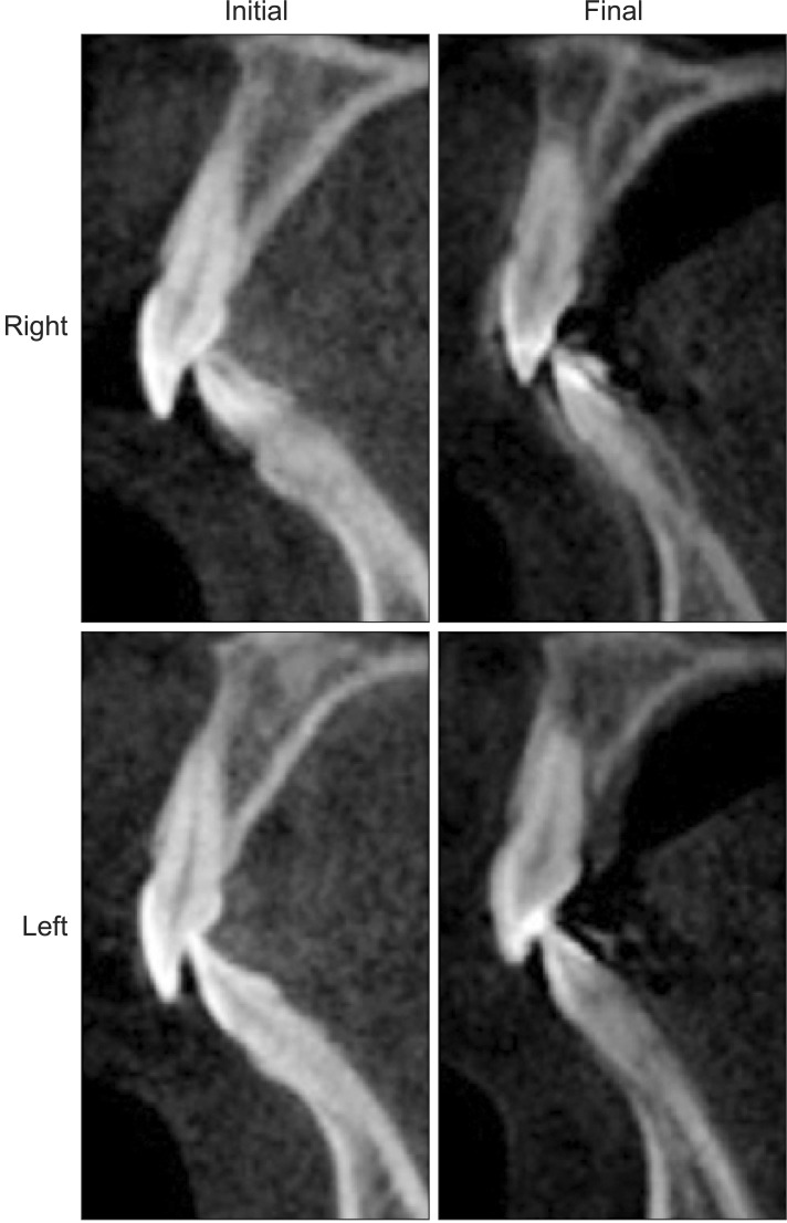
Figure 13 Changes in the central incisors and the surrounding alveolar housing. Mild root blunting is noted, but the incisors are within their biological boundaries after treatment.
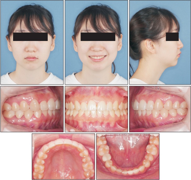
Figure 14 Postretention facial and intraoral photographs.
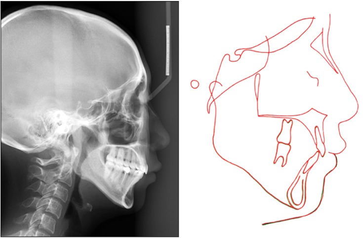
Figure 15 Cephalometric superimposition at debonding and after 15 months of follow-up. Black, final; red, retention.
Cited by 1 articles
-
Total arch distalization with interproximal stripping in a patient with severe crowding
Min-Ho Jung
Korean J Orthod. 2019;49(3):194-201. doi: 10.4041/kjod.2019.49.3.194.
Reference
-
1. Garber DA, Salama MA. The aesthetic smile: diagnosis and treatment. Periodontol 2000. 1996; 11:18–28. PMID: 9567953.
Article2. Chang CA, Fields HW Jr, Beck FM, Springer NC, Firestone AR, Rosenstiel S, et al. Smile esthetics from patients' perspectives for faces of varying attractiveness. Am J Orthod Dentofacial Orthop. 2011; 140:e171–e180. PMID: 21967955.
Article3. Hunt O, Johnston C, Hepper P, Burden D, Stevenson M. The influence of maxillary gingival exposure on dental attractiveness ratings. Eur J Orthod. 2002; 24:199–204. PMID: 12001557.
Article4. An SM, Choi SY, Chung YW, Jang TH, Kang KH. Comparing esthetic smile perceptions among laypersons with and without orthodontic treatment experience and dentists. Korean J Orthod. 2014; 44:294–303. PMID: 25473645.
Article5. Hwang WS, Hur MS, Hu KS, Song WC, Koh KS, Baik HS, et al. Surface anatomy of the lip elevator muscles for the treatment of gummy smile using botulinum toxin. Angle Orthod. 2009; 79:70–77. PMID: 19123705.
Article6. Kim SJ, Kim JW, Choi TH, Lee KJ. Combined use of miniscrews and continuous arch for intrusive root movement of incisors in Class II division 2 with gummy smile. Angle Orthod. 2014; 84:910–918. PMID: 24512532.
Article7. Proffit WR, Phillips C, Turvey TA. Stability following superior repositioning of the maxilla by LeFort I osteotomy. Am J Orthod Dentofacial Orthop. 1987; 92:151–161. PMID: 3475970.
Article8. Parekh SM, Fields HW, Beck M, Rosenstiel S. Attractiveness of variations in the smile arc and buccal corridor space as judged by orthodontists and laymen. Angle Orthod. 2006; 76:557–563. PMID: 16808559.9. Baek MS, Choi YJ, Yu HS, Lee KJ, Kwak J, Park YC. Long-term stability of anterior open-bite treatment by intrusion of maxillary posterior teeth. Am J Orthod Dentofacial Orthop. 2010; 138:396.e1–396.e9. discussion 396-8. PMID: 20889043.
Article10. Kuroda S, Sakai Y, Tamamura N, Deguchi T, Takano-Yamamoto T. Treatment of severe anterior open bite with skeletal anchorage in adults: Comparison with orthognathic surgery outcomes. Am J Orthod Dentofacial Orthop. 2007; 132:599–605. PMID: 18005833.
Article11. Deguchi T, Kurosaka H, Oikawa H, Kuroda S, Takahashi I, Yamashiro T, et al. Comparison of orthodontic treatment outcomes in adults with skeletal open bite between conventional edgewise treatment and implant-anchored orthodontics. Am J Orthod Dentofacial Orthop. 2011; 139(4 Suppl):S60–S68. PMID: 21435540.
Article12. Arai C, Choi JW, Nakaoka K, Hamada Y, Nakamura Y. Management of open bite that developed during treatment for internal derangement and osteoarthritis of the temporomandibular joint. Korean J Orthod. 2015; 45:136–145. PMID: 26023542.
Article13. Bechtold TE, Kim JW, Choi TH, Park YC, Lee KJ. Distalization pattern of the maxillary arch depending on the number of orthodontic miniscrews. Angle Orthod. 2013; 83:266–273. PMID: 22970751.
Article14. Sohn BW, Lee JG, Kim HS, Kim HS. Semi-longitudinal study of growth and development of children aged 6 to 17 Part I: Growth and development of arch form. Korean J Orthod. 1996; 26:225–239.15. Burstone CJ, James RB, Legan H, Murphy GA, Norton LA. Cephalometrics for orthognathic surgery. J Oral Surg. 1978; 36:269–277. PMID: 273073.16. Ioi H, Kang S, Shimomura T, Kim SS, Park SB, Son WS, et al. Effects of buccal corridors on smile esthetics in Japanese and Korean orthodontists and orthodontic patients. Am J Orthod Dentofacial Orthop. 2012; 142:459–465. PMID: 22999668.
Article17. Park YC. A morphologic study on straight wire bracket for Korean. Korean J Orthod. 1991; 21:481–493.18. Zachrisson BU. Buccal uprighting of canines and premolars for improved smile esthetics and stability. World J Orthod. 2006; 7:406–412. PMID: 17190235.19. Schendel SA, Carlotti AE Jr. Variations of total vertical maxillary excess. J Oral Maxillofac Surg. 1985; 43:590–596. PMID: 3859608.
Article20. Choi YJ, Lee JS, Cha JY, Park YC. Total distalization of the maxillary arch in a patient with skeletal Class II malocclusion. Am J Orthod Dentofacial Orthop. 2011; 139:823–833. PMID: 21640890.
Article21. Kim SJ, Choi TH, Baik HS, Park YC, Lee KJ. Mandibular posterior anatomic limit for molar distalization. Am J Orthod Dentofacial Orthop. 2014; 146:190–197. PMID: 25085302.
Article22. Lee KJ, Joo E, Kim KD, Lee JS, Park YC, Yu HS. Computed tomographic analysis of tooth-bearing alveolar bone for orthodontic miniscrew placement. Am J Orthod Dentofacial Orthop. 2009; 135:486–494. PMID: 19361735.
Article23. Jeong GM, Sung SJ, Lee KJ, Chun YS, Mo SS. Finite-element investigation of the center of resistance of the maxillary dentition. Korean J Orthod. 2009; 39:83–94.
Article24. Sung EH, Kim SJ, Chun YS, Park YC, Yu HS, Lee KJ. Distalization pattern of whole maxillary dentition according to force application points. Korean J Orthod. 2015; 45:20–28. PMID: 25667914.
Article25. Shafiee R, Korn EL, Pearson H, Boyd RL, Baumrind S. Evaluation of facial attractiveness from end-of-treatment facial photographs. Am J Orthod Dentofacial Orthop. 2008; 133:500–508. PMID: 18405813.
Article26. Yamada K, Kuroda S, Deguchi T, Takano-Yamamoto T, Yamashiro T. Distal movement of maxillary molars using miniscrew anchorage in the buccal interradicular region. Angle Orthod. 2009; 79:78–84. PMID: 19123698.
Article27. Proffit WR. The third stage of comprehensive treatment: finishing. In : Proffit WR, Fields HW, Sarver DV, editors. Contemporary orthodontics. 5th ed. St. Louis: Elsevier Health Sciences;2014. p. 596–597.28. Murakami T, Yokota S, Takahama Y. Periodontal changes after experimentally induced intrusion of the upper incisors in Macaca fuscata monkeys. Am J Orthod Dentofacial Orthop. 1989; 95:115–126. PMID: 2916468.
Article29. Melsen B. Tissue reaction following application of extrusive and intrusive forces to teeth in adult monkeys. Am J Orthod. 1986; 89:469–475. PMID: 3459360.
Article30. Han G, Huang S, Von den Hoff JW, Zeng X, Kuijpers-Jagtman AM. Root resorption after orthodontic intrusion and extrusion: an intraindividual study. Angle Orthod. 2005; 75:912–918. PMID: 16448231.
- Full Text Links
-
- Actions
-
Cited
- CITED
-
- Close
- Share
-
- Similar articles
-
- Distalization with a modified C-palatal plate for severe upper crowding and a missing lower incisor
- Anterior esthetic restoration using DSD (digital smile design) for a patient with congenital missing tooth of maxillary central incisor
- Change in arch width in extraction vs nonextraction treatment
- Orthodontic treatment of gummy smile by maxillary total intrusion with a midpalatal absolute anchorage system
- Immediate changes in the mandibular dentition after maxillary molar distalization using headgear
