Virtual otoscopy for evaluating the inner ear with a fluid-filled tympanic cavity in dogs
- Affiliations
-
- 1Department of Radiological Technology, Daegu Health College, Daegu 702-722, Korea.
- 2College of Veterinary Medicine, Chonbuk National University, Jeonju 561-756, Korea. kclee@chonbuk.ac.kr
Abstract
- The feasibility of virtual otoscopy (VO) imaging was evaluated in five dogs with experimentally induced otitis media, two control dogs, and two canine patients with otitis media. VO images of the tympanic cavity and ossicles were generated with commercially available software using raw computed tomography (CT) data. Eight out of 10 ears inoculated with pathogen exhibited obvious clinical signs associated with otitis externa. CT images revealed soft tissue density material occupying the tympanic bulla compatible with otitis media in three dogs with experimentally induced otitis media and two patients. No remarkable features were observed on the radiographs. Four different VO views (ear canal, tympanic bulla, eustachian tube, and ossicular chain) were created. Promontory, cochlea window, tympanic, and septum bulla as well as ossicles were easily and clearly distinguished except for the incus and stapes of the clinical patients. VO images were not more suitable than images created with conventional CT for accurately diagnosing otitis media in this study. However, it appears that VO could be more feasible for assessing the complex structure of the inner ear in dogs with fluid-filled tympanic cavities since fluid accumulation within the tympanic bulla did not affect the evaluation of bony tissue in the middle ear on VO images.
Keyword
MeSH Terms
Figure
-

Fig. 1 Transverse computed tomography (CT) image of the temporal bone with the dog in a dorsal recumbent position. Fluid opacity was seen around the tympanic bulla. The arrow within the circle shows the virtual otoscopy (VO) direction of Fig. 4. EC: ear canal, TB: tympanic bulla, M: malleus, I: incus, CW: cochlear window, D: dorsal, V: ventral, MED: medial, LAT: lateral.

Fig. 2 Transverse CT image of the temporal bone with the dog in a ventral recumbent position. Fluid level in the ventral aspect was observed in the tympanic bulla. The arrow within the circle shows the VO direction of Fig. 5.

Fig. 3 Transverse CT image of the temporal bone in a canine patient with otitis media placed in a ventral recumbent position. A fluid-filled tympanic bulla and an absence of air density in the ear canal were observed. The arrow within the circle shows the VO direction of Fig. 6.
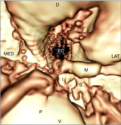
Fig. 4 VO view from the tympanic bulla toward the auditory tube. Fluid opacity did not interfere with identifying the internal structures of the middle ear. S: stapes, P: promontory.

Fig. 5 VO view from the tympanic bulla toward the auditory tube.
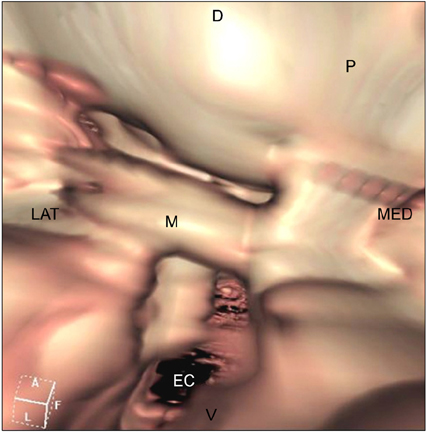
Fig. 6 VO view from the tympanic bulla toward the auditory tube in the same dog shown in Fig. 5.
Reference
-
1. Allgoewer I, Lucas S, Schmitz SA. Magnetic resonance imaging of the normal and diseased feline middle ear. Vet Radiol Ultrasound. 2000. 41:413–418.
Article2. Barthez PY, Koblik PD, Hornof WJ, Wisner ER, Seibert JA. Apparent wall thickening in fluid filled versus air filled tympanic bulla in computed tomography. Vet Radiol Ultrasound. 1996. 37:95–98.
Article3. Boor S, Maurer J, Mann W, Stoeter P. Virtual endoscopy of the inner ear and the auditory canal. Neuroradiology. 2000. 42:543–547.
Article4. Cole LK, Samii VF. Contrast-enhanced computed tomographic imaging of the auditory tube in mesaticephalic dogs. Vet Radiol Ultrasound. 2007. 48:125–128.
Article5. Dvir E, Kirberger RM, Terblanche AG. Magnetic resonance imaging of otitis media in a dog. Vet Radiol Ultrasound. 2000. 41:46–49.
Article6. Eom K, Kwak H, Kang H, Park S, Lee H, Kang H, Kwon J, Kim I, Kim N, Lee K. Virtual CT otoscopy of the middle ear and ossicles in dogs. Vet Radiol Ultrasound. 2008. 49:545–550.
Article7. Fabinyi B, Klug C. A minimally invasive technique for endoscopic middle ear surgery. Eur Arch Otorhinolaryngol. 1997. 254:Suppl 1. S53–S54.
Article8. Frankenthaler R, Moharir V, Kikinis R, van Kipshagen P, Jolesz F, Umans C, Fried MP. Virtual otoscopy. Otolaryngol Clin North Am. 1998. 31:383–392.
Article9. Garosi LS, Dennis R, Schwarz T. Review of diagnostic imaging of ear diseases in the dog and cat. Vet Radiol Ultrasound. 2003. 44:137–146.
Article10. Griffiths LG, Sullivan M, O'Neill T, Reid SWJ. Ultrasonography versus radiography for detection of fluid in the canine tympanic bulla. Vet Radiol Ultrasound. 2003. 44:210–213.
Article11. Imori T, Matsuda H, Tohjo M, Kamata Y. An experimental production of suppurative otitis media in dog, and a trial to evaluate the therapeutic effect of cefmetazole on this otitis media. Jpn J Antibiot. 1982. 35:2277–2287.12. Karhuketo TS, Dastidar PS, Ryymin PS, Laasonen EM, Puhakka HJ. Virtual endoscopy imaging of the middle ear cavity and ossicles. Eur Arch Otorhinolaryngol. 2002. 259:77–83.
Article13. Kneissl S, Probst A, Konar M. Low-field magnetic resonance imaging of the canine middle and inner ear. Vet Radiol Ultrasound. 2004. 45:520–522.
Article14. Lee J, Eom K, Seong Y, Lee H, Park J, Lee J, Jang K, Lee K, Oh T, Lee S, Yoon J, Lee H, Choi H, Lee Y, Chang D. Ultrasonographic evaluation of the external ear canal and tympanic membrane in dogs. Vet Radiol Ultrasound. 2006. 47:94–98.
Article15. Love NE, Kramer RW, Spodnick GJ, Thrall DE. Radiographic and computed tomographic evaluation of otitis media in the dog. Vet Radiol Ultrasound. 1995. 36:375–379.
Article16. Neri E, Caramella D, Panconi M, Berrettini S, Sellari Franceschini S, Forli F, Bartolozzi C. Virtual endoscopy of the middle ear. Eur Radiol. 2001. 11:41–49.
Article17. Owen MC, Lamb CR, Lu D, Targett MP. Material in the middle ear of dogs having magnetic resonance imaging for investigation of neurologic signs. Vet Radiol Ultrasound. 2004. 45:149–155.
Article18. Pandey AK, Bapuraj JR, Gupta AK, Khandelwal N. Is there a role for virtual otoscopy in the preoperative assessment of the ossicular chain in chronic suppurative otitis media? Comparison of HRCT and virtual otoscopy with surgical findings. Eur Radiol. 2009. 19:1408–1416.
Article19. Rawlings CA. Diagnostic rigid endoscopy: otoscopy, rhinoscopy, and cystoscopy. Vet Clin North Am Small Anim Pract. 2009. 39:849–868.
Article20. Rodt T, Bartling S, Schmidt AM, Weber BP, Lenarz T, Becker H. Virtual endoscopy of the middle ear: experimental and clinical results of a standardised approach using multi-slice helical computed tomography. Eur Radiol. 2002. 12:1684–1692.
Article21. Rodt T, Ratiu P, Becker H, Bartling S, Kacher DF, Anderson M, Jolesz FA, Kikinis R. 3D visualisation of the middle ear and adjacent structures using reconstructed multi-slice CT datasets, correlating 3D images and virtual endoscopy to the 2D cross-sectional images. Neuroradiology. 2002. 44:783–790.
Article22. Rohleder JJ, Jones JC, Duncan RB, Larson MM, Waldron DL, Tromblee T. Comparative performance of radiography and computed tomography in the diagnosis of middle ear disease in 31 dogs. Vet Radiol Ultrasound. 2006. 47:45–52.
Article23. Shell LG. Otitis media and otitis interna. Etiology, diagnosis, and medical management. Vet Clin North Am Small Anim Pract. 1988. 18:885–899.24. Silverstein H, Jackson LE. Wiet RJ, editor. Office-based minor surgery: otoendoscopy and inner ear perfusion. Ear and Temporal Bone Surgery: Minimizing Risks and Complications. 2006. 1st ed. New York: Thieme Medical Publishers;275–284.25. Trojanowska A, Czekajska-Chehab E, Trojanowski P, Olszanski W, Klatka J, Drop A, Golabek W. Comparison of multidetector row CT cross-sectional source images with multiplanar 2D-, 3D- reconstructions and virtual endoscopy in assessment of the middle ear. J Neuroradiol. 2006. 33:277–278.
Article26. Yamada K, Morimoto M, Kishimoto M, Wisner ER. Virtual endoscopy of dogs using multi-detector row CT. Vet Radiol Ultrasound. 2007. 48:318–322.
Article
- Full Text Links
-
- Actions
-
Cited
- CITED
-
- Close
- Share
-
- Similar articles
-
- Referred Otalgia Induced by a Large Tonsillolith
- Aberrant Internal Carotid Artery in the Middle Ear
- Tympanic Membrane Perforation Due to Metal Spark in a Welder
- Tympanosclerosis of the Middle Ear: Radiologic-Surgical Correlation
- Clinical Usefulness of Temperature of Tympanic Membrane in Diagnosing Unilateral Acute Suppurative Otitis Media
