Focal Cerebral Ischemia Reduces Protein Phosphatase 2A Subunit B Expression in Brain Tissue and HT22 Cells
- Affiliations
-
- 1Department of Anatomy, College of Veterinary Medicine, Research Institute of Life Sciences, Gyeongsang National University, Jinju, Republic of Korea. pokoh@gnu.ac.kr
Abstract
- Protein phosphatase 2A (PP2A) is a serine and threonine protein phosphatase that regulates cell cycle progression and apoptosis. PP2A is composed of various subunits. Among these subunits, subunit B plays an important role in the modulation of PP2A function in the brain. This study investigated PP2A subunit B expression levels after neuronal cell injury. Middle cerebral artery occlusions (MCAO) were surgically induced in adult male rats to induce focal cerebral ischemic injury, and brain tissues were collected 24 h after MCAO. A proteomic approach revealed reduction of PP2A subunit B protein spots in MCAO-operated animals in comparison to sham-operated animals. Western blot analysis confirmed that MCAO induces reductions in PP2A subunit B levels. Moreover, glutamate exposure induces neuronal cell death and leads to reductions of PP2A subunit B levels in a hippocampal-derived cell line. This study demonstrated the decrease of PP2A subunit B in ischemic neuronal cell injury. These results suggest that the decrease of PP2A subunit B after ischemic brain injury can mediate neuronal cell death.
MeSH Terms
Figure
-
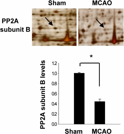
Figure 1 Protein phosphatase 2A (PP2A) subunit B protein spots identified by MALDI-TOF. Arrows indicate the protein spots. The intensity of spots was measured using PDQuest software. The ratio of intensity is described as spots intensity of middle cerebral artery-occluded (MCAO) animal to spots intensity of sham-operated animal. Data are shown as mean±SEM. *P<0.05 (vs. Sham).
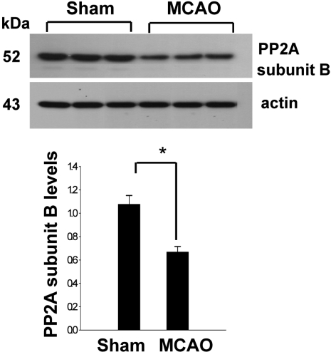
Figure 2 Western blot analysis of protein phosphatase 2A (PP2A) subunit B in the cerebral cortex from sham-operated and middle cerebral artery-occluded (MCAO) animals. Each lane represents an individual experimental animal. Densitometric analysis of PP2A levels is represented as intensity of PP2A to intensity of actin. Data are shown as mean±SEM. *P<0.05 (vs. Sham).
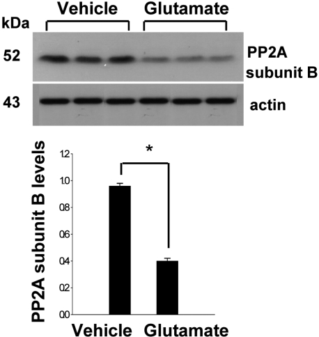
Figure 3 Western blot analysis of protein phosphatase 2A (PP2A) subunit B in HT22 cells. Glutamate (5 mM) or vehicle was exposed to HT22 cells for 24 h. Each lane represents an individual experimental animal. Densitometric analysis of PP2A levels is represented as intensity of PP2A to intensity of actin. Data are shown as mean±SEM. *P<0.05 (vs. Vehicle).
Reference
-
1. Ferrer I, Planas AM. Signaling of cell death and cell survival following focal cerebral ischemia: life and death struggle inthe penumbra. J Neuropathol Exp Neurol. 2003; 62(4):329–339. PMID: 12722825.2. Gong CK, Lidsky T, Wegiel J, Zuck L, Grundke-Iqbal I, Iqbal K. Phosphorylation of microtubule-associated protein tau is regulated by protein phosphatase 2A in mammalian brain. Implications for neurofibrillary degeneration in Alzheimer's disease. J Biol Chem. 2000; 275(8):5535–5544. PMID: 10681533.3. Janssens V, Goris J. Protein phosphatase 2A: a highly regulated family of serine/threonine phosphatases implicated in cell growth and signalling. Biochem J. 2001; 353(Pt 3):417–439. PMID: 11171037.
Article4. Koh PO. 17Beta-estradiol prevents the glutamate-induced decrease of Akt and its downstream targets in HT22 cells. J Vet Med Sci. 2007; 69(3):285–288. PMID: 17409645.
Article5. Koh PO. Proteomic analysis of focal cerebral ischemic injury in male rats. J Vet Med Sci. 2010; 72(2):181–185. PMID: 19942814.
Article6. Li Y, Chopp M, Powers C, Jiang N. Apoptosis and protein expression after focal cerebral ischemia in rat. Brain Res. 1997; 765(2):301–312. PMID: 9313903.
Article7. Liu R, Wang JZ. Protein phosphatase 2A in Alzheimer's disease. Pathophysiology. 2009; 16(4):273–277. PMID: 19278841.
Article8. Longa EZ, Weinstein PR, Carlson S, Cummins R. Reversible middle cerebral artery occlusion without craniectomy in rats. Stroke. 1989; 20(1):84–91. PMID: 2643202.
Article9. Maher P, Davis JB. The role of monoamine metabolism inoxidative glutamate toxicity. J Neurosci. 1996; 16(20):6394–6401. PMID: 8815918.10. Millward TA, Zolnierowicz S, Hemmings BA. Regulation of protein kinase cascades by protein phosphatase 2A. Trends Biochem Sci. 1999; 24(5):186–191. PMID: 10322434.
Article11. Siesjö BK. Mechanisms of ischemic brain damage. Crit Care Med. 1988; 16(10):954–963. PMID: 3048896.12. Sontag E, Nunbhakdi-Craig V, Lee G, Bloom GS, Mumby MC. Regulation of the phosphorylation state and microtubule-binding activity of Tau by protein phosphatase 2A. Neuron. 1996; 17(6):1201–1207. PMID: 8982166.
Article13. Sontag E. Protein phosphatase 2A: the Trojan Horse of cellular signaling. Cell Signal. 2001; 13(1):7–16. PMID: 11257442.14. Stone SR, Hofsteenge J, Hemmings BA. Molecular cloning of cDNAs encoding two isoforms of the catalytic subunit ofprotein phosphatase 2A. Biochemistry. 1987; 26(23):7215–7220. PMID: 2827745.
- Full Text Links
-
- Actions
-
Cited
- CITED
-
- Close
- Share
-
- Similar articles
-
- Curcumin treatment recovery the decrease of protein phosphatase 2A subunit B induced by focal cerebral ischemia in Sprague-Dawley rats
- Epigallocatechin gallate restores the reduction of protein phosphatase 2 A subunit B caused by middle cerebral artery occlusion
- Focal Cerebral Ischemia Induces Decrease of Astrocytic Phosphoprotein PEA-15 in Brain Tissue and HT22 Cells
- Decrease of protein phosphatase 2A subunit B by glutamate exposure in the cerebral cortex of neonatal rats
- Chlorogenic acid regulates the expression of protein phosphatase 2A subunit B in the cerebral cortex of a rat stroke model and glutamate-exposed neurons
