Mapping of the Faded (fe) Gene to a Region between D10mit191 and D10mit44 on Mouse Chromosome 10
- Affiliations
-
- 1Department of Medical Genetics, College of Medicine, Hallym University, Chuncheon, Republic of Korea. jgsuh@hallym.ac.kr
- 2Institute of Natural Medicine, Hallym University, Chuncheon, Republic of Korea.
Abstract
- The faded mouse is a coat color mutant that shows faded coat color and age-related loss of pigmentation. This mutation is transmitted by an autosomal recessive gene with 100% penetrance. In the present study, we carried out linkage analysis of the faded (fe) gene using intra-specific backcross panels. Affected faded mice were carefully confirmed by their faded coat color at about 4 weeks of age. In the intra-specific backcross between faded and CBA mice (n=198), the fe gene was mapped to a region 2.1 cM distal to D10mit191. Therefore, the gene order was defined as follows: centromere-D10mit51 (12.4+/-2.4 cM)-D10mit191 (2.1+/-1.0 cM)-fe-D10mit44 (13.3+/-2.4 cM)-D10mit42 (14.4+/-2.5 cM). This linkage map of the fe locus will provide a good entry point to isolate the fe gene. Since the faded mouse has pigmentary abnormalities, this mutant may be a useful model for studies of pigmentary abnormalities in humans.
Keyword
MeSH Terms
Figure
-
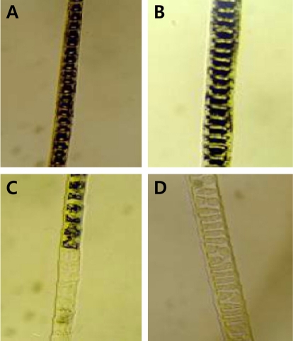
Figure 1 Age-related pigment change of affected N2 progenies. A: 1 week, B: 2 weeks, C: 4 weeks, D: 28 weeks.
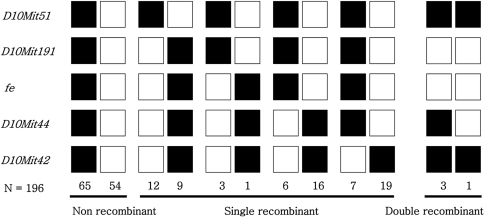
Figure 2 Haplotype analysis of an intraspecific backcross between fe and CBA/J mice [(C57BL/6J-fe/fe×CBA/J)F1×C57BL/6J-fe/fe]. Each column represents a chromosomal haplotype identified in the backcross and inherited from the F1 parent. The number of individuals is listed beneath each column. The black boxes represent fe alleles, and the white boxes represent CBA/J alleles as determined by SSLP scoring with markers indicated at the left.
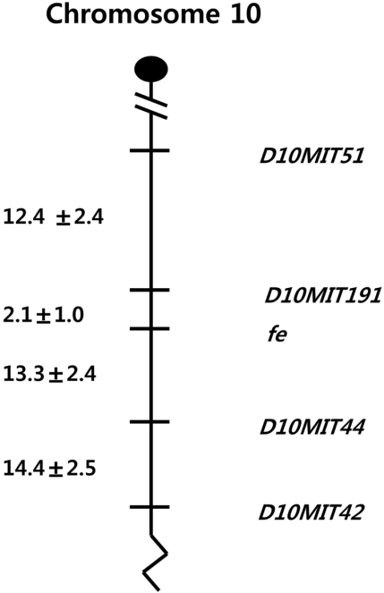
Figure 3 Genetic linkage maps of chromosome 10 as derived from an intra backcross panel [(C57BL/6J-fe/fe×CBA/J)F1×C57BL/6J-fe/fe] generated in this study. Distances between intervals are shown in centimorgans±standard error to the left of the map.
Reference
-
1. Brilliant MH. The mouse p (pink-eyed dilution) and human P genes, oculocutaneous albinism type 2 (OCA2), and melanosomal pH. Pigment Cell Res. 2001; 14(2):86–93. PMID: 11310796.2. Cable J, Jackson IJ, Steel KP. Light (Blt), a mutation that causes melanocyte death, affects stria vascularis function in the mouse inner ear. Pigment Cell Res. 1993; 6:215–225. PMID: 8248019.
Article3. Edery P, Attie T, Amiel J, Pelet A, Eng C, Hofstra RM, Martelli H, Bidaud C, Munnich A, Lyonnet S. Mutation of the endothelin-3 gene in the Waardenburg-Hirschsprung disease (Shah-Waardenburg syndrome). Nat Genet. 1996; 12(4):442–444. PMID: 8630502.
Article4. Fukuta K, Imamura K, Goto N. Pink-eyed dilution, a coat color mutation in the Japanese field vole (Microtus montebelli). Jikken Dobutsu. 1991; 40(3):375–379. PMID: 1915604.5. Green EL. Linkage, recombination and mapping. Genetics and probability in animal breeding experiments. 1981. Oxford University Press: New York;p. 77–133.
Article6. Grant SG, O'Dell TJ, Karl KA, Stein PL, Soriano P, Kandel ER. Impaired long-term potentiation, spatial learning and hippocampal development in fyn mutant mice. Science. 1992; 258(5090):1903–1910. PMID: 1361685.7. Gariepy CE, Williams SC, Richardson JA, Hammer RE, Yanagisawa M. Transgenic expression of the endothelin-B receptor prevents congenital intestinal aganglionosis in a rat model of Hirschsprung disease. J Clin Invest. 1998; 102(6):1092–1101. PMID: 9739043.
Article8. Johnson R, Jackson IJ. Light is a dominant mouse mutation resulting in premature cell death. Nat Genet. 1992; 1(3):226–229. PMID: 1303241.
Article9. Kobayashi T, Vicira WD, Pottcrt B, Sakai C, Imokawa G, Hearing VJ. Light Modulation of melanogenic protein expression during the switch from eu- to phaeomelanogenesis. J Cell Sci. 1995; 108:2301–2309. PMID: 7673350.10. Kunisada T, Yoshida H, Yamazaki H, Miyamoto A, Hemmi H, Nishimura E, Shultz LD, Nishikawa S, Hayashi S. Transgene expression of steel factor in the basal layer of epidermis promotes survival, proliferation, differentiation and migration of melanocyte precursors. Development. 1998; 125(15):2915–2923. PMID: 9655813.
Article11. Lamoreux ML. Strain-specific white-spotting patterns in laboratory mice. Pigment Cell Res. 1999; 12(6):383–390. PMID: 10614578.
Article12. Lehman AL, Silvers WK, Puri N, Wakamatsu K, Ito S, Brilliant MH. The underwhite (uw) locus acts autonomously and reduces the production of melanin. J Invest Dermatol. 2000; 115:601–606. PMID: 10998130.
Article13. Marks PW, Bandura JL, Shieh DB, Foernzler D, Beier DR, Kwiatkowski DJ. The spontaneous coat color mutant white nose (wn) maps to murine Chromosome 15. Mamm Genome. 1999; 10(7):750–752. PMID: 10384053.14. Oh YS, Tomita T. Linkage of faded gene (fe) to chromosome 6 of the mouse. Jikken Dobutsu. 1987; 36(1):73–77. PMID: 3816991.15. Oh YS, Tomita T, Kondo K. Faded, a mutation in the KSB strain of mouse which shows age-related pigment change. Jikken Dobutsu. 1986; 35(2):131–138. PMID: 3732403.
Article16. O'Reilly FM, Brat DJ, McAlpine BE, Grossniklaus HE, Folpe AL, Arbiser JL. Microphthalmia transcription factor immunohistochemistry: a useful diagnostic marker in the diagnosis and detection of cutaneous melanoma, sentinel lymph node metastases, and extracutaneous melanocytic neoplasms. J Am Acad Dermatol. 2001; 45(3):414–419. PMID: 11511840.17. Pastural E, Barrat FJ, Dufourcq-Lagelouse R, Certain S, Sanal O, Jabado N, Seger R, Criscelli C, Fischer A, de Saint Basile G. Griscelli disease maps to chromosome 15q21 and is associated with mutations in the Myosin Va gene. Nature Genet. 1997; 16:289–292. PMID: 9207796.18. Richards KA, Fukai K, Oiso N, Paller AS. A novel KIT mutation results in piebaldism with progressive depigmentation. J Am Acad Dermatol. 2001; 44(2):288–292. PMID: 11174389.
Article19. Straile WZ, Chase HB, Apenault C. Growth and differentiation of hair follicles between periods of activity and quiescence. J Exp Zool. 1961; 148:205–222. PMID: 13917600.
Article20. Silvers WK. The coat colors of mice. 1979. New York: Springer-Verlag;p. 1–5.21. Slominski A, Paus R. Melanogenesis is coupled to murine angen: toward new concepts for the role of melanocytes and the regulation of melanogensis in hair growth. J Invest Dermatol. 1993; 101:90s–97s. PMID: 8326158.22. Slominski A, Paus R, Plonka P, Chakraborty A, Maurer M, Pruski D, Lukiewicz S. Melanogenesis during the anagen-catagentelogen transformation of the murine hair cycle. J Invest Dermatol. 1994; 102:862–862. PMID: 8006449.
Article23. Stumpo DJ, Bock CB, Tuttle JS, Blackshear PJ. MARCKS deficiency in mice leads to abnormal brain development and perinatal death. Proc Natl Acad Sci. 1995; 92(4):944–948. PMID: 7862670.
Article24. Suh JG, Yamanishi T, Matsui K, Tanaka K, Wada K. Mapping of the gracile axonal dystrophy (gad) gene to a region between D5mit197 and D5mit113 on proximal mouse chromosome 5. Genomics. 1995; 27:549–551. PMID: 7558041.25. Shanske A, Ferreira JC, Leonard JC, Fuller P, Marion RW. Hirschsprung disease in an infant with a contiguous gene syndrome of chromosome 13. Am J Med Genet. 2001; 102(3):231–236. PMID: 11484199.
Article26. Tomita Y, Miyamura Y, Kono M, Nakamura R, Matsunaga J. Molecular bases of congenital hypopigmentary disorders in humans and oculocutaneous albinism 1 in Japan. Pigment Cell Res. 2000; 13(Suppl 8):130–134. PMID: 11041370.
Article27. Tobin DJ, Paus R. Graying: gerontobiology of the hair follicle pigmentary unit. Exp Gerontol. 2001; 36(1):29–54. PMID: 11162910.
Article28. Toyofuku K, Wada I, Valencia JC, Kushimoto T, Ferrans VJ, Hearing VJ. Oculocutaneous albinism types 1 and 3 are ER retention diseases: mutation of tyrosinase or Tyrp1 can affect the processing of both mutant and wild-type proteins. FASEB J. 2001; 15(12):2149–2161. PMID: 11641241.
Article29. Voisey J, Box NF, van Daal A. A polymorphism study of the human Agouti gene and its association with MC1R. Pigment Cell Res. 2001; 14(4):264–267. PMID: 11549109.
Article30. Wang C, Kim E, Attaie A, Smith TN, Wilcox ER, Lalwani AK. A PAX3 polymorphism (T315K) in a family exhibiting Waardenburg Syndrome type 2. Mol Cell Probes. 1998; 12(1):55–57. PMID: 9584079.31. Wada A, Okumoto M, Tsudzuki M. Tawny: a novel light coat color mutation found in a wild population of Mus musculus molossinus, a new allele at the melanocortin 1 receptor (Mc1r) locus. Exp Anim. 1999; 48(2):73–78. PMID: 10374067.
- Full Text Links
-
- Actions
-
Cited
- CITED
-
- Close
- Share
-
- Similar articles
-
- A high resolution genetic mapping of the faded (fe) gene to a region between D10mit156 and D10mit193 on mouse chromosome 10
- Characterization of a Novel Gene in the Extended MHC Region of Mouse, NG29/Cd320, a Homolog of the Human CD320
- Cloning of mouse AQP-CD gene
- A Case of 46 , XX Male
- Detection of chromosome aberrations in interphase nuclei using fluorescence in situ hybridization technique
