Primary malignant melanoma of cervix and vagina
- Affiliations
-
- 1Department of Obstetrics and Gynecology, Institute of Women's Medical Life Science, Yonsei University College of Medicine, Seoul, Korea. SAN1@yuhs.ac
- 2Department of Obstetrics and Gynecology, Samsung Medical Center, Sungkyunkwan University School of Medicine, Seoul, Korea.
- 3Department of Pathology, Samsung Medical Center, Sungkyunkwan University School of Medicine, Seoul, Korea.
- KMID: 2391695
- DOI: http://doi.org/10.5468/ogs.2016.59.5.415
Abstract
- Primary malignant melanoma (MM) accounts for 1% of all cancers, and only 3% to 7% of these tumors occur in the female genital tract. Data are limited with respect to the basis for treatment recommendations because of the rarity of MM. The overall prognosis of melanomas of the female genital tract is very poor. Two cases of MM of the female genital tract are presented. The first case is of a 70-year-old female patient who complained of left thigh pain and underwent magnetic resonance imaging that showed cervical cancer with involvement of the vagina, bladder, and parametrium, in addition to multiple bony metastases of the proximal femur, acetabulum, and both iliac bones. The second case is of a 35-year-old female patient who suffered from vaginal bleeding for 5 months, and she was diagnosed as having primary vaginal melanoma. The patient underwent radical surgery and two additional surgeries because of recurrence of cancer in both inguinal areas. After surgery, the patient received adjuvant immunotherapy, radiation therapy, and chemotherapy. In both the aforementioned cases, the pathologic diagnosis was made after immunohistochemical analysis, i.e., the tumor cells were stained with HMB-45 and S100, and were found to be positive for both immunostains.
Keyword
MeSH Terms
Figure
-
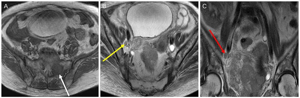
Fig. 1 Magnetic resonance imaging findings. (A) About 8.6×7.3-cm enhancing cervical mass shows an invasion to the vaginal, bladder, and parametrium. It demonstrates mixed intermediate to subtle high signal intensity on T1WI. The white arrow indicates left sacral metastasis. (B) Right obturator lymph node metastasis is noted (yellow arrow). It shows heterogeneous intermediate to subtle high signal intensity on T2WI. (C) Right obturator lymph node metastasis and diffuse pelvic bone metastasis are noted (red arrow). It shows heterogeneous enhancement after contrast administration.
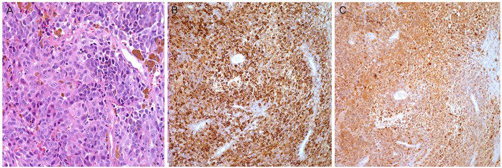
Fig. 2 (A) Highly atypical sheets of malignant cells reveal sufficient granular cytoplasm and prominent eosinophilic nucleoli. The atypical cells show diverse morphology from the spindle to epithelioid cells, and mixed in brownish pigments and abundant blood vessels (H&E, ×400 ). (B) Immunohistochemical stains for HMB45 (×200) were done. Antibodies against HMB 45 (melanosome) disclose diffused positive reaction against tumor cells. (C) Immunohistochemical stains for S100 (×100) were done. Antibodies against S100 protein disclose diffused positive reaction against tumor cells.
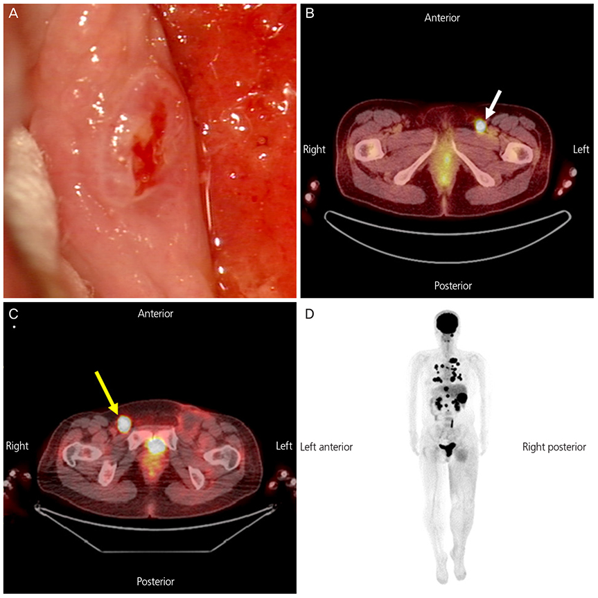
Fig. 3 (A) Colposcopic findings of primary malignant melanoma at vagina. It shows pinkish ulcerative lesion at lower vagina. (B) positron emission tomography-computed tomography (PET-CT) findings of recurrence of vaginal melanoma at left inguinal lymph node (white arrow). (C) PET-CT findings of recurrence of vaginal melanoma at right inguinal node (yellow arrow). (D) PET-CT findings of metastasis to multiple organs 37 months after the initial diagnosis, following three times of surgery and adjuvant therapy.
Reference
-
1. Xia L, Han D, Yang W, Li J, Chuang L, Wu X. Primary malignant melanoma of the vagina: a retrospective clinicopathologic study of 44 cases. Int J Gynecol Cancer. 2014; 24:149–155.2. Pusceddu S, Bajetta E, Buzzoni R, Carcangiu ML, Platania M, Del Vecchio M, et al. Primary uterine cervix melanoma resembling malignant peripheral nerve sheath tumor: a case report. Int J Gynecol Pathol. 2008; 27:596–600.3. Seifried S, Haydu LE, Quinn MJ, Scolyer RA, Stretch JR, Thompson JF. Melanoma of the vulva and vagina: principles of staging and their relevance to management based on a clinicopathologic analysis of 85 cases. Ann Surg Oncol. 2015; 22:1959–1966.4. Chang AE, Karnell LH, Menck HR. The National Cancer Data Base report on cutaneous and noncutaneous melanoma: a summary of 84,836 cases from the past decade. The American College of Surgeons Commission on Cancer and the American Cancer Society. Cancer. 1998; 83:1664–1678.5. Cantuaria G, Angioli R, Fernandez-Abril A, Penalver M. Primary malignant melanoma of the uterine cervix: case report and review of the literature. Prim Care Update Ob Gyns. 1998; 5:159–160.6. Janco JM, Markovic SN, Weaver AL, Cliby WA. Vulvar and vaginal melanoma: case series and review of current management options including neoadjuvant chemotherapy. Gynecol Oncol. 2013; 129:533–537.7. Mordel N, Mor-Yosef S, Ben-Baruch N, Anteby SO. Malignant melanoma of the uterine cervix: case report and review of the literature. Gynecol Oncol. 1989; 32:375–380.8. Singh K, DiSilvestro PA, Lawrence WD, Quddus MR. An isolated metastasis from clear cell renal cell carcinoma to the uterus: a case report and review of literature. Int J Gynecol Pathol. 2016; 35:419–422.9. Lokadasan R, Ratheesan K, Sukumaran R, Nair SP. Metastatic lobular carcinoma of breast mimics primary cervix carcinoma: two case reports and a review of the literature. Ecancermedicalscience. 2015; 9:571.10. Clark KC, Butz WR, Hapke MR. Primary malignant melanoma of the uterine cervix: case report with world literature review. Int J Gynecol Pathol. 1999; 18:265–273.11. Morrow CP, DiSaia PJ. Malignant melanoma of the female genitalia: a clinical analysis. Obstet Gynecol Surv. 1976; 31:233–271.12. Frumovitz M, Etchepareborda M, Sun CC, Soliman PT, Eifel PJ, Levenback CF, et al. Primary malignant melanoma of the vagina. Obstet Gynecol. 2010; 116:1358–1365.13. Pinedo F, Ingelmo JM, Miranda P, Garzon A, Lopez JI. Primary malignant melanoma of the uterine cervix: case report and review of the literature. Gynecol Obstet Invest. 1991; 31:121–124.14. Buchanan DJ, Schlaerth J, Kurosaki T. Primary vaginal melanoma: thirteen-year disease-free survival after wide local excision and review of recent literature. Am J Obstet Gynecol. 1998; 178:1177–1184.15. Frumovitz M, Gayed IW, Jhingran A, Euscher ED, Coleman RL, Ramirez PT, et al. Lymphatic mapping and sentinel lymph node detection in women with vaginal cancer. Gynecol Oncol. 2008; 108:478–481.
- Full Text Links
-
- Actions
-
Cited
- CITED
-
- Close
- Share
-
- Similar articles
-
- A Case of Primary Malignant Melanoma of the Vagina: Trial of a Wide Local Excision of Vagina and Rectum
- Malignant melanoma of the vagina: CT and MR findings
- Cytologic Features of Papnicolaou Smears of Malignant Melanoma Arising in Vagina: A Cese Report
- A Case of Primary Malignant Melanoma of the Vagina
- A Case of Primary Malignant Melanoma of Cervix and Vagina
