Feasibility of Ultrasound Guided Atlanto-occipital Joint Injection
- Affiliations
-
- 1Department of Rehabilitation Medicine, Yeouido St. Mary's Hospital, Seoul 150-713, Korea. shewry@hanmail.net
- 2Department of Anatomy, The Catholic University of Korea, College of Medicine, Seoul 150-713, Korea.
Abstract
OBJECTIVE
To evaluate the feasibility of ultrasound guided atlanto-occipital joint injection. METHOD: Six atlanto-occipital joints of three cadavers were examined. Cadavers were placed in prone position with their head slightly rotated towards the contra-lateral side. The atlanto-occipital joint was initially identified with a longitudinal ultrasound scan at the midline between occipital protuberance and mastoid process. Contrast media 0.5cc was injected into the atlanto-occipital joint using an in-plane needle approach under ultrasound guide. The location of the needle tip and spreading pattern of the contrast was confirmed by fluoroscopic evaluation.
RESULTS
After ultrasound guided atlanto-occipital joint injection, spreading of the contrast media into the joint was seen in all the injected joints in the anterior-posterior fluoroscopic view.
CONCLUSION
The ultrasound guided atlanto-occipital injection is feasible. The ultrasound guided injection by Doppler examination can provide a safer approach to the atlanto-occipital joint.
MeSH Terms
Figure
-
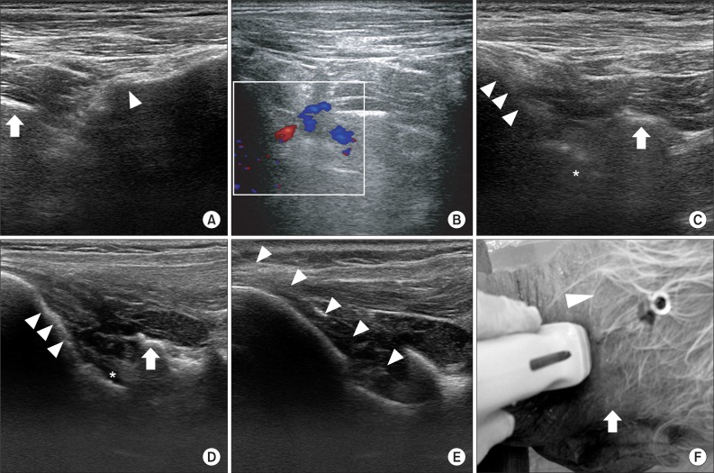
Fig. 1 Ultrasound guided atlanto-occipital joint injection. (A) In ultrasound view of living body at the longitudinal scan below the mastoid process, C1 transverse process (arrow) and C2-3 facet joint (arrowhead) are shown. (B) Doppler examination in living body. Vertebral artery is visualized between C1 transverse process and occiput. (C) In ultrasound view of living body with a longitudinal scan at the mid-point between mastoid process and occipital protuberance, atlanto-occipital joint (asterix) is seen between C1 transverse process (arrow) and occiput (arrowhead). (D) In ultrasound view of cadaver, same as C, atlanto-occipital joint (asterix), C1 transverse process (arrow), and occiput (arrowhead) are shown. The atlanto-occipital joint is more clearly visualized than in living body. (E) In real time ultrasound guided injection, needle (arrowhead) is placed at the atlanto-occipital joint. (F) Needle insertion point is the mid-point between occipital protuberance (arrowhead) and mastoid process (arrow).
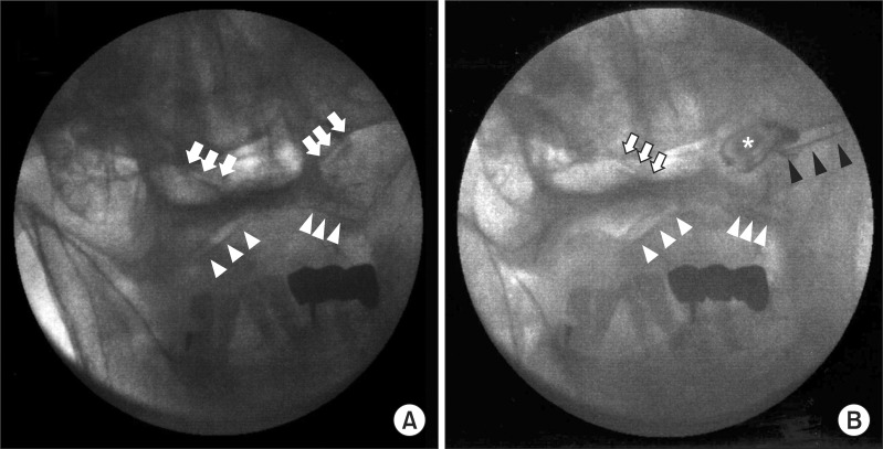
Fig. 2 Anterior-posterior fluoroscopic view of atlanto-occipital joint. (A) Before contrast media injection, bilateral atlanto-occipital joints (arrow) and atlanto-axial joints (arrowhead) are seen. (B) After contrast media injection, needle placement (black arrow) and spreading to the atlanto-occipital joint (asterix) are clearly visualized.
Reference
-
1. Ogoke BA. The management of the atlanto-occipital and atlanto-axial joint pain. Pain Physician. 2000; 3:289–293. PMID: 16906186.
Article2. Meleger AL, Krivickas LS. Neck and back pain: musculoskeletal disorders. Neurol Clin. 2007; 25:419–438. PMID: 17445737.
Article3. Halla JT, Hardin JG, Vitek J, Alarcon GS. Involvement of the cervical spine in rheumatoid arthritis. Arthritis Rheum. 1989; 32:652–659. PMID: 2655607.
Article4. Busch E, Wilson PR. Atlanto-occipital and atlanto-axial injections in the treatment of headache and neck pain. Reg Anesth. 1989; 14:45.5. Dreyfuss P, Rogers J, Dreyer S, Fletcher D. Atlanto-occipital joint pain. A report of three cases and description of an intraarticular joint block technique. Reg Anesth. 1994; 19:344–351. PMID: 7848935.6. Menezes AH, Traynelis VC. Anatomy and biomechanics of normal craniovertebral junction (a) and biomechanics of stabilization (b). Childs Nerv Syst. 2008; 24:1091–1100. PMID: 18389261.
Article7. Tokuda K, Miyasaka K, Abe H, Abe S, Takei H, Sugimoto S, Tsuru M. Anomalous atlantoaxial portions of vertebral and posterior inferior cerebellar arteries. Neuroradiology. 1985; 27:410–413. PMID: 4058734.
Article8. Uchino A, Saito N, Watadani T, Okada Y, Kozawa E, Nishi N, Mizukoshi W, Inoue K, Nakajima R, Takahashi M. Vertebral artery variations at the C1-2 level diagnosed by magnetic resonance angiography. Neuroradiology. 2012; 54:19–23. PMID: 21340577.
Article9. Yoshihara H, Kepler C, Hasegawa K, Rawlins BA. Surgical treatment for atlantooccipital osteoarthritis: a case report of two patients. Eur Spine J. 2011; 20(Suppl 2):S243–S247. PMID: 21125298.
Article10. Valdez DC, Johnson RG. Role of technetium-99m planar bone scanning in the evaluation of low back pain. Skeletal Radiol. 1994; 23:91–97. PMID: 8191307.
Article11. Falco FJ, Erhart S, Wargo BW, Bryce DA, Atluri S, Datta S, Hayek SM. Systematic review of diagnostic utility and therapeutic effectiveness of cervical facet joint interventions. Pain Physician. 2009; 12:323–344. PMID: 19305483.12. Manchikanti L, Singh V, Falco FJ, Cash KM, Fellows B. Cervical medial branch blocks for chronic cervical facet joint pain: a randomized, double-blind, controlled trial with one-year follow-up. Spine. 2008; 33:1813–1820. PMID: 18670333.13. Pekkafahli MZ, Kiralp MZ, Basekim CC, Silit E, Mutlu H, Ozturk E, Kizilkaya E, Dursun H. Sacroiliac joint injections performed with sonographic guidance. J Ultrasound Med. 2003; 22:553–559. PMID: 12795552.14. Greher M, Scharbert G, Kamolz LP, Beck H, Gustorff B, Kirchmair L, Kapral S. Ultrasound-guided lumbar facet nerve block: a sonoanatomic study of a new methodologic approach. Anesthesiology. 2004; 100:1242–1248. PMID: 15114223.15. Galiano K, Obwegeser AA, Bodner G, Freund MC, Gruber H, Maurer H, Schatzer R, Fiegele T, Ploner F. Ultrasound-guided facet joint injections in the middle to lower cervical spine : a CT-controlled sonoanatomic study. Clin J Pain. 2006; 22:538–543. PMID: 16788340.
- Full Text Links
-
- Actions
-
Cited
- CITED
-
- Close
- Share
-
- Similar articles
-
- Atlanto-Occipital Dislocation: A Case Report
- Feasibility of Ultrasound-Guided Intraarticular Contrast Injection for MR Arthrography
- Traumatic Atlanto-Occipital Rotatory Posterior Dislocation Combined with Atlanto-Axial Rotatory Subluxation: A Case Report
- Essential Clinical Tips about Ultrasound Guided Cervical Intervention
- Cervical Myelopathy Secondary to Atlanto-occipital Assimilation: The Usefulness of the Simple Decompressive Surgery
