Recurrent osteochondroma of the mandibular condyle: A case report
- Affiliations
-
- 1Department of Oral and Maxillofacial Radiology, School of Dentistry, Kyungpook National University, Daegu, Korea. syan@knu.ac.kr
- 2Department of Oral and Maxillofacial Surgery, School of Dentistry, Kyungpook National University, Daegu, Korea.
- 3Department of Oral and Maxillofacial Radiology, School of Dentistry, Chonnam National University, Gwangju, Korea.
- KMID: 2391408
- DOI: http://doi.org/10.5624/isd.2017.47.1.57
Abstract
- A 21-year-old woman presented with facial asymmetry. Crepitus and clicking of the temporomandibular joint were noted. The midline deviated 5.5 mm to the left, and secondary malocclusion was observed. Panoramic and cone-beam computed tomographic images showed an irregular and exophytic bony mass on the anteromedial surface of the right mandibular condyle. A 3-phase bone scan revealed increased tracer uptake on the affected side. The lesion was treated with excision and reshaping under the diagnosis of osteochondroma confirmed by a histopathological examination. The lesion recurred after 3 years, and the patient underwent condylectomy. Mandibular condylar osteochondroma is often resected because it causes functional and aesthetic problems, but it rarely recurs. To the best of our knowledge, only 2 cases of recurrent osteochondromas of the mandibular condyle have been reported previously. Surgical treatment of the osteochondroma should be performed considering the possibility of recurrence, and long-term follow-up is recommended.
MeSH Terms
Figure
-

Fig. 1 A. Preoperative photograph shows facial asymmetry with chin deviation to the left side. B. Intraoral photograph reveals midline deviation to the left (arrow) and secondary malocclusion in the closed position.
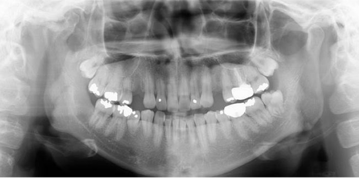
Fig. 2 Panoramic radiograph reveals an irregular and large bony mass extending from the anterior portion of the right condylar head.
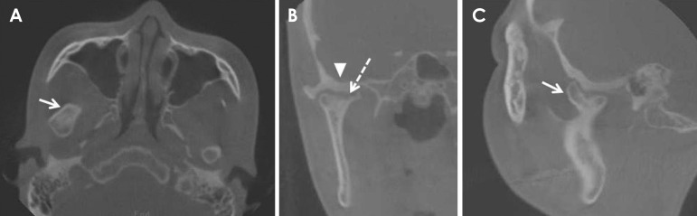
Fig. 3 A-C. Cone-beam computed tomography (CBCT) images demonstrate the well-defined margin of the radiopaque mass on the anteromedial surface of the condylar neck (arrow) with a focal erosive change (dotted arrow). Depression and cortical thickenings of the skull base (arrowhead) can also be seen.
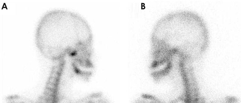
Fig. 4 A. 99mTc-hydroxydiphosphonate bone scan, with a lateral spot view, showing an abnormal tracer uptake in the right condylar head. B. No significant tracer avidity is seen on the left side.
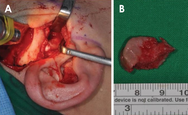
Fig. 5 A. A pre-auricular approach was used for exposing the condyle. B. The surgical specimen was approximately 20 mm in length.
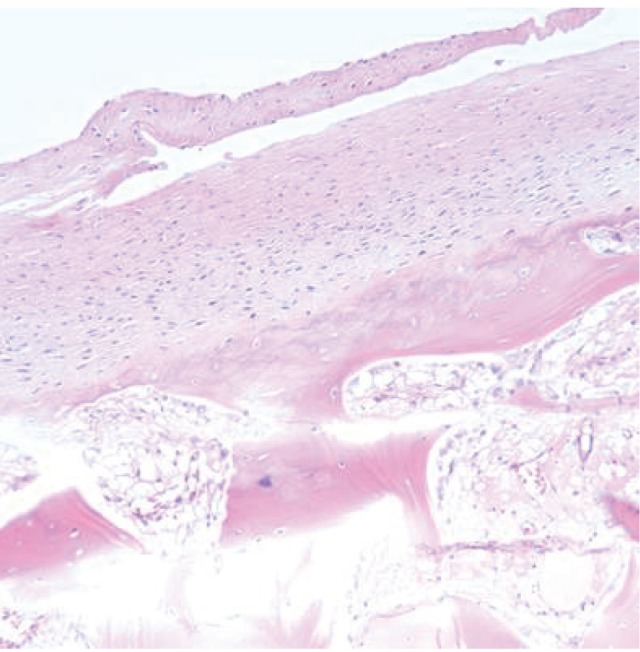
Fig. 6 Osteochondroma showing a fibrous perichondrium and chondroid matrix with chondrocytes in the lacuna. The cartilaginous tissue is seen blending with the cancellous bone (hematoxylin and eosin, original magnification: ×200).
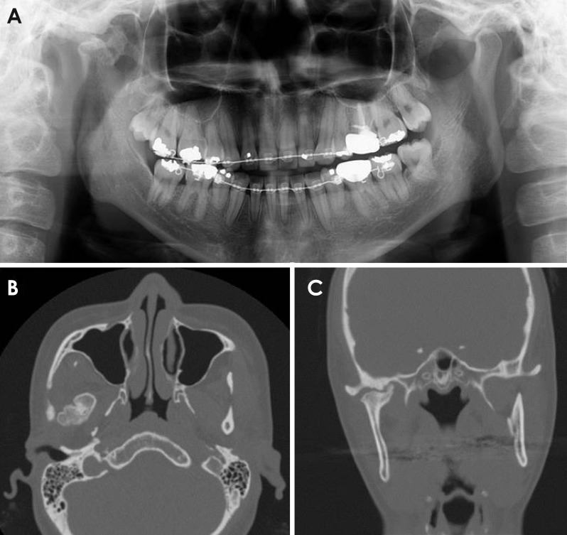
Fig. 7 A. Panoramic radiograph of recurred osteochondroma shows the exophytic mass on the right condyle. B-C. An axial and coronal computed tomograph shows a similar pattern of bony outgrowth on the affected side.
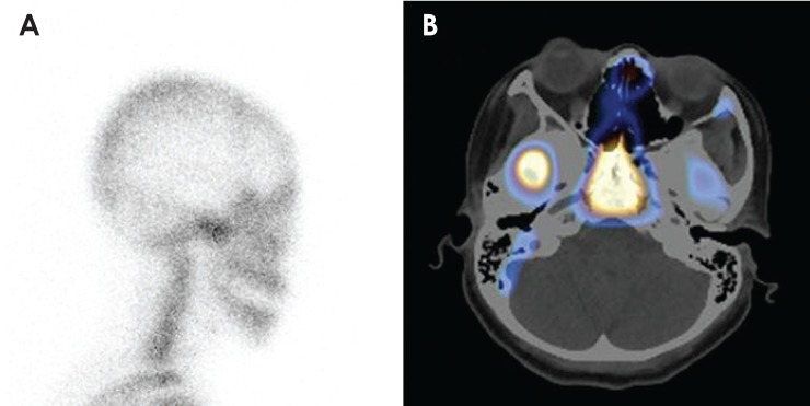
Fig. 8 A. Bone scan reveals increased tracer uptake in the recurrence area. B. Fused single-photon emission computed tomography (SPECT)/CT shows intense tracer uptake in the affected condylar area.
Reference
-
1. Zhou Q, Yang C, Chen MJ. Osteochondroma of bilateral mandibular condyle: a case report. Int J Clin Exp Med. 2015; 8:2989–2992. PMID: 25932269.2. Karras SC, Wolford LM, Cottrell DA. Concurrent osteochondroma of the mandibular condyle and ipsilateral cranial base resulting in temporomandibular joint ankylosis: report of a case and review of the literature. J Oral Maxillofac Surg. 1996; 54:640–646. PMID: 8632254.3. Aydin MA, Küçükçelebi A, Sayilkan S, Celebioğlu S. Osteochondroma of the mandibular condyle: report of 2 cases treated with conservative surgery. J Oral Maxillofac Surg. 2001; 59:1082–1089. PMID: 11526583.4. Arora P, Deora SS, Kiran S, Bargale SD. Osteochondroma of condyle: case discussion and review of treatment modalities. BMJ Case Rep. 2014; 2014:pii: bcr2013200759.
Article5. Wolford LM, Mehra P, Franco P. Use of conservative condylectomy for treatment of osteochondroma of the mandibular condyle. J Oral Maxillofac Surg. 2002; 60:262–268. PMID: 11887135.
Article6. Vezeau PJ, Fridrich KL, Vincent SD. Osteochondroma of the mandibular condyle: literature review and report of two atypical cases. J Oral Maxillofac Surg. 1995; 53:954–963. PMID: 7629631.7. Peroz I. Osteochondroma of the condyle: case report with 15 years of follow-up. Int J Oral Maxillofac Surg. 2016; 45:1120–1122. PMID: 27156430.
Article8. Peroz I, Scholman H, Hell B. Osteochondroma of the mandibular condyle: a case report. Int J Oral Maxillofac Surg. 2002; 31:455–456. PMID: 12361086.
Article9. Zhang J, Wang H, Li X, Li W, Wu H, Miao J, et al. Osteochondromas of the mandibular condyle: variance in radiographic appearance on panoramic radiographs. Dentomaxillofac Radiol. 2008; 37:154–160. PMID: 18316507.
Article10. Avinash KR, Rajagopal KV, Ramakrishnaiah RH, Carnelio S, Mahmood NS. Computed tomographic features of mandibular osteochondroma. Dentomaxillofac Radiol. 2007; 36:434–436. PMID: 17881606.
Article11. Kondoh T, Seto K, Kobayashi K. Osteoma of the mandibular condyle: report of a case with a review of the literature. J Oral Maxillofac Surg. 1998; 56:972–979. PMID: 9710193.
Article12. Chandu A, Spencer JA, Dyson DP. Chondroma of the mandibular condyle: an example of a rare tumour. Dentomaxillofac Radiol. 1997; 26:242–245. PMID: 9442616.
Article13. Dhirawani RB, Anand K, Lalwani G, Pathak S, Thakkar B. True chondroma of the mandibular condyle: a rare case. Ann Maxillofac Surg. 2014; 4:220–223. PMID: 25593880.
Article
- Full Text Links
-
- Actions
-
Cited
- CITED
-
- Close
- Share
-
- Similar articles
-
- The Osteochondroma of the Mandibular Condyle: report of a case
- Surgical Treatment of Osteochondroma on the Mandibular Condyle through Intraoral Approach: Case Report
- Osteochonrdoma Of The Mandibular Condyle: A Case Report
- Conservative resection of osteochondroma on mandibular condyle: A case report
- Giant osteochondroma of the parapharyngeal space: a case report
