Intramuscular hemangioma in buccal cheek: a case report
- Affiliations
-
- 1Department of Oral and Maxillofacial Surgery, College of Dentistry, Dankook University, Cheonan, Korea. kimchoms@dankook.ac.kr
- KMID: 2391357
- DOI: http://doi.org/10.5125/jkaoms.2017.43.4.262
Abstract
- Hemangioma is the most common benign tumor of a vascular origin, and is characterized by the abnormal proliferation of blood vessels. Intramuscular hemangioma (IMH) usually involves the skeletal muscles of the trunk or limbs, but rarely occurs in the head and neck region. This case report presents a patient with IMH showing multiple phleboliths in the buccal cheek. A 13-year-old boy was referred for the evaluation and management of painful swelling of the left cheek that had gradually increased in size over a 6 year duration. The examination revealed a palpable firm mass. Reddish-blue buccal mucosa color was observed with an aciniform shape. Preoperative magnetic resonance imaging (MRI) showed a vascular tumor in the left side adjacent to the buccinator and depressor orbicularis oris muscles. Surgical resection under general anesthesia was performed via the intraoral approach. The mass and phleboliths were extracted successfully. A histopathological examination confirmed the diagnosis of IMH. In conclusion, clinicians should be aware of the possibility of IMH in cases of a palpable mass with multiple nodules deep within the muscle in the buccal cheek. Among the several diagnostic tools, MRI provides essential information on the extent and surrounding anatomy of IMH.
MeSH Terms
Figure
-
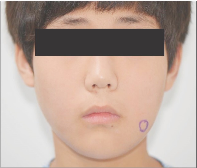
Fig. 1 Extraoral clinical photo.
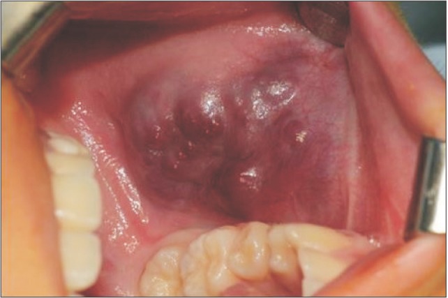
Fig. 2 Intraoral clinical photo.
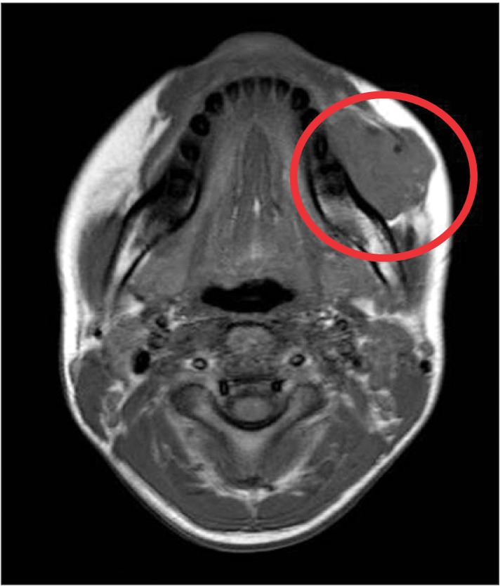
Fig. 3 Axial view on preoperative magnetic resonance imaging (T1-weighted). A circle shows isointense mass measuring 4×3 cm in size with well-contoured margin in the left abutting on the mandible, buccinator muscle, depressor orbicularis oris muscle, and subcutaneous fat layer.
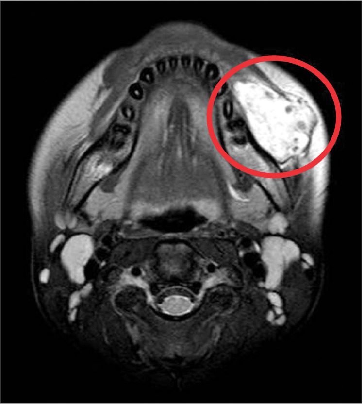
Fig. 4 Axial view on preoperative magnetic resonance imaging (T2-weighted). A circle shows hyperintense mass with hypointense spaces suggesting phleboliths.
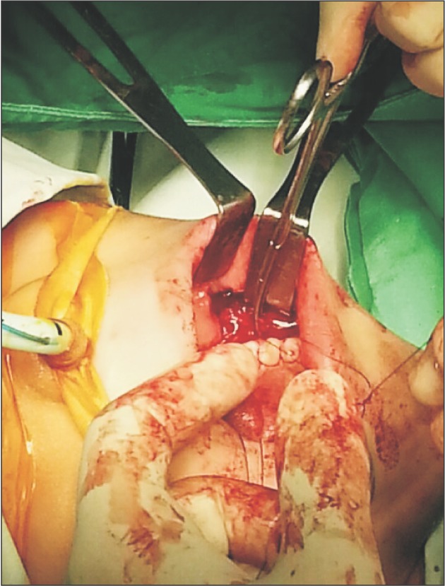
Fig. 5 Local bleeding control by ligation of feeding vessel.
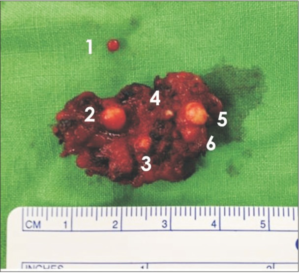
Fig. 6 Extracted mass with several phleboliths.
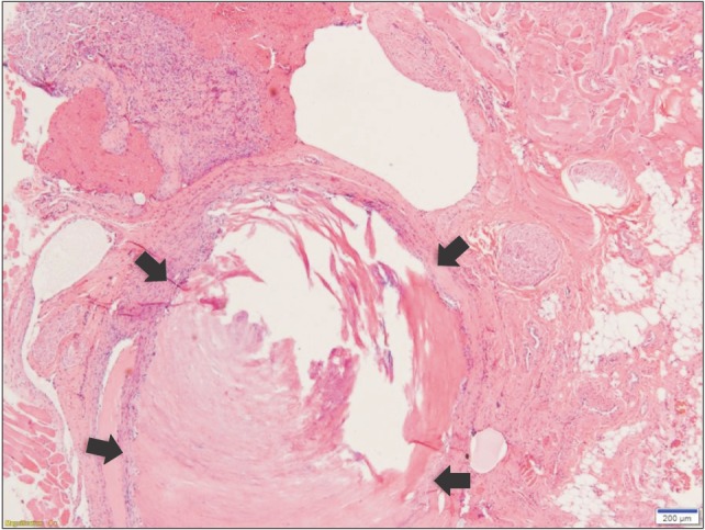
Fig. 7 Cut surface of phlebolith (H&E staining, ×40). The phlebolith in the arrows appears concentric structure suggesting occurrence of sclerosis and the midportion looks more deep purple color because of minute calcification.
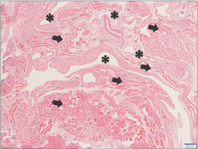
Fig. 8 Histopathologic examination of intramuscular hemangioma specimen (H&E staining, ×40). Asterisks indicate blood vessels sprouted into the muscles and multiple arrows point out the various patterns of muscle arrangement.
Reference
-
1. Altuğ HA, Büyüksoy V, Okçu KM, Doğan N. Hemangiomas of the head and neck with phleboliths: clinical features, diagnostic imaging, and treatment of 3 cases. Oral Surg Oral Med Oral Pathol Oral Radiol Endod. 2007; 103:e60–e64.
Article2. Mandel L, Surattanont F. Clinical and imaging diagnoses of intramuscular hemangiomas: the wattle sign and case reports. J Oral Maxillofac Surg. 2004; 62:754–758. PMID: 15170294.
Article3. Rossiter JL, Hendrix RA, Tom LW, Potsic WP. Intramuscular hemangioma of the head and neck. Otolaryngol Head Neck Surg. 1993; 108:18–26. PMID: 8437870.
Article4. Ingalls GK, Bonnington GJ, Sisk AL. Intramuscular hemangioma of the mentalis muscle. Oral Surg Oral Med Oral Pathol. 1985; 60:476–481. PMID: 3864109.
Article5. Kanaya H, Saito Y, Gama N, Konno W, Hirabayashi H, Haruna S. Intramuscular hemangioma of masseter muscle with prominent formation of phleboliths: a case report. Auris Nasus Larynx. 2008; 35:587–591. PMID: 18207684.
Article6. Odabasi AO, Metin KK, Mutlu C, Başak S, Erpek G. Intramuscular hemangioma of the masseter muscle. Eur Arch Otorhinolaryngol. 1999; 256:366–369. PMID: 10473832.
Article7. Righini CA, Berta E, Atallah I. Intramuscular cavernous hemangioma arising from the masseter muscle. Eur Ann Otorhinolaryngol Head Neck Dis. 2014; 131:57–59. PMID: 23845293.
Article8. Saeed WR, Kolhe PS, Smith FW, Murray GI. The ‘turkey wattle’ sign revisited: diagnosing parotid vascular malformations in the adult. Br J Plast Surg. 1997; 50:43–46. PMID: 9038514.
Article9. Biller HF, Krespi YP, Som PM. Combined therapy for vascular lesions of the head and neck with intra-arterial embolization and surgical excision. Otolaryngol Head Neck Surg. 1982; 90:37–47. PMID: 6806755.
Article10. Yonetsu K, Nakayama E, Yuasa K, Kanda S, Ozeki S, Shinohara M. Imaging findings of some buccomasseteric masses. Oral Surg Oral Med Oral Pathol Oral Radiol Endod. 1998; 86:755–759. PMID: 9868738.
Article11. Clemis JD, Briggs DR, Changus GW. Intramuscular hemangioma in the head and neck. Can J Otolaryngol. 1975; 4:339–346. PMID: 1139430.12. Demir Z, Oktem F, Celebioğlu S. Rare case of intramasseteric cavernous hemangioma in a three-year-old boy: a diagnostic dilemma. Ann Otol Rhinol Laryngol. 2004; 113:455–458. PMID: 15224828.
Article13. Yonetsu K, Nakayama E, Miwa K, Tanaka T, Araki K, Kanda S, et al. Magnetic resonance imaging of oral and maxillofacial angiomas. Oral Surg Oral Med Oral Pathol. 1993; 76:783–789. PMID: 8284086.
Article14. Osada K, Yoshihara T, Itoh M. Intramasseter hemangiomas: a case report. J Otolaryngol. 2000; 29:166–169. PMID: 10883831.15. Zengin AZ, Celenk P, Sumer AP. Intramuscular hemangioma presenting with multiple phleboliths: a case report. Oral Surg Oral Med Oral Pathol Oral Radiol. 2013; 115:e32–e36.
Article16. Ichimura K, Nibu K, Tanaka T. Essentials of surgical treatment for intramasseteric hemangioma. Eur Arch Otorhinolaryngol. 1995; 252:125–129. PMID: 7662343.
Article17. Addante RR, Donovan MG. Right facial mass. J Oral Maxillofac Surg. 1994; 52:1061–1065. PMID: 8089793.
Article18. Allen PW, Enzinger FM. Hemangioma of skeletal muscle. An analysis of 89 cases. Cancer. 1972; 29:8–22. PMID: 5061701.
- Full Text Links
-
- Actions
-
Cited
- CITED
-
- Close
- Share
-
- Similar articles
-
- Incidentally Found Intramuscular Hemangioma, Mimicking Traumatic Hematoma after Military Training: A Case Report
- Intramuscular hemangioma formation in the masseter muscle: a case report
- Intramuscular Hemangioma of the Mentalis Muscle: A Case Report
- Cavernous Hemangioma of the Masseter Muscle
- Multiple Myeloma In Buccal Cheek Mucosa: Report Of A Case And Review Of Literatures
