Recording natural head position using an accelerometer and reconstruction from computed tomographic images
- Affiliations
-
- 1Department of Oral and Maxillofacial Surgery, College of Dentistry, Wonkwang University, Iksan, Korea. kkhoms@hanmail.net
- 2Department of Orthodontics, College of Dentistry, Wonkwang University, Iksan, Korea.
- 3College of Dentistry, Graduate School, Wonkwang University, Iksan, Korea.
- KMID: 2391356
- DOI: http://doi.org/10.5125/jkaoms.2017.43.4.256
Abstract
OBJECTIVES
The concept of natural head position (NHP) was first introduced by Broca in 1862, and was described as a person's stable physiologic position "when a man is standing and his visual axis is horizontal." NHP has been used routinely for clinical examination; however, a patient's head position is random during cone-beam computed tomography (CBCT) acquisition. To solve this problem, we developed an accelerometer to record patients' NHP and reproduce them for CBCT images. In this study, we also tested the accuracy and reproducibility of our accelerometer.
MATERIALS AND METHODS
A total of 15 subjects participated in this study. We invented an accelerometer that measured acceleration on three axes and that could record roll and pitch calculations. Recorded roll and pitch data for each NHP were applied to a reoriented virtual image using three-dimensional (3D) imaging software. The data between the 3D models and the clinical photos were statistically analyzed side by side. Paired t-tests were used to statistically analyze the measurements.
RESULTS
The average difference in the angles between the clinical photograph and the 3D model was 0.04° for roll and 0.29° for pitch. The paired ttests for the roll data (P=0.781) and the pitch data (P=0.169) showed no significant difference between the clinical photographs and the 3D model (P>0.05).
CONCLUSION
By overcoming the limitations of previous NHP-recording techniques, our new method can accurately record patient NHP in a time-efficient manner. Our method can also accurately transfer the NHP to a 3D virtual model.
MeSH Terms
Figure
-
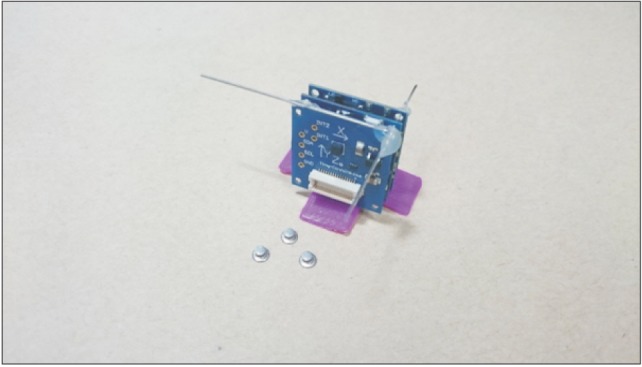
Fig. 1 The accelerometer and three lingual buttons. We made a small accelerometer (3×3×2 cm, 60 g) using the TinyDuino Platform (TinyCircuits, USA) with a Nordic BLE nRF8001 bluetooth 4.0 chip and Bosch BMA250 3-axis accelerator chip on it. Roll and pitch are calculated per 0.5 second with inverse trigonometric function.
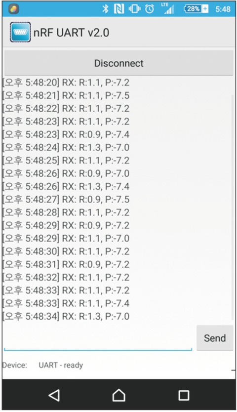
Fig. 2 Measuring roll and pitch with an application. Roll and pitch data of the device were transmitted to android smartphone application (nRF UART v2.0). Averaged last five data were used in this study.
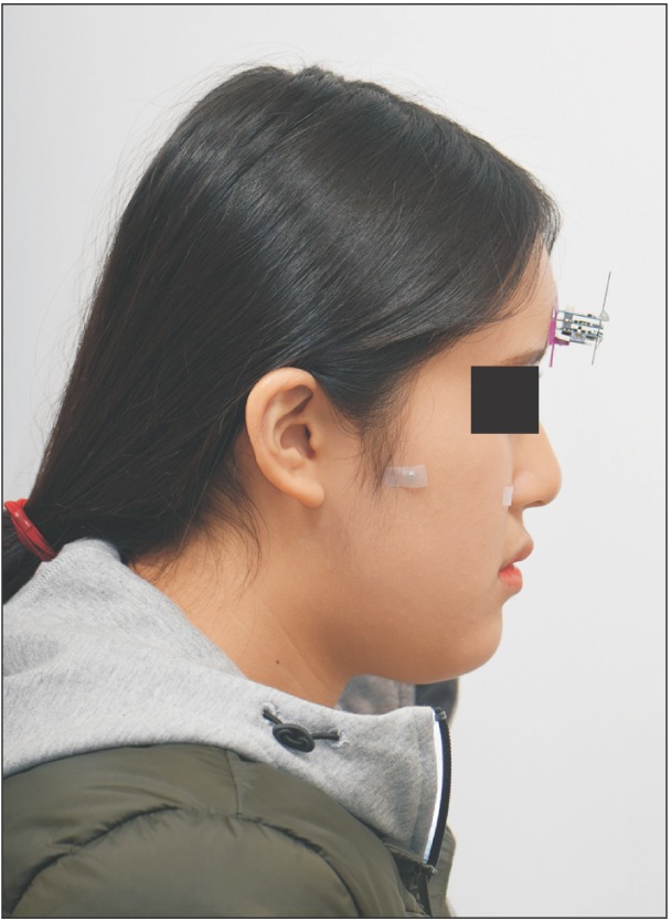
Fig. 3 Clinical photo (lateral side). Two lingual buttons, one on the periauricular and another on the right zygomatic area are shown. To compare the pitch value, we made a line connecting those buttons, and the angle was calculated.
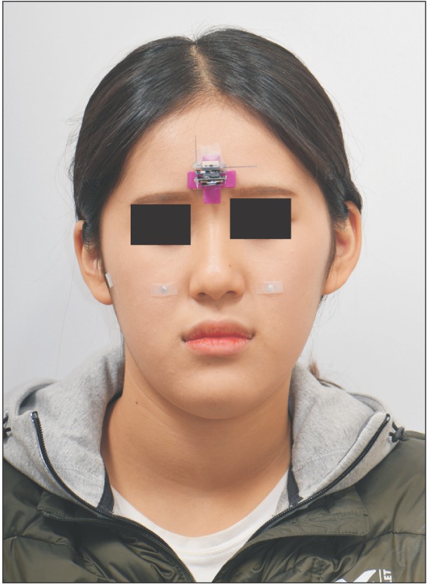
Fig. 4 Clinical photo (frontal side). Two lingual buttons on the bilateral zygomatic area are shown. To compare the roll value, we made a line between those buttons, and the angle was measured.
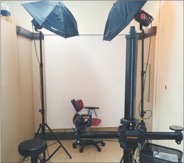
Fig. 5 Clinical photo room. The mirror on the wall was used to orient patient's natural head position by mirror-guided technique. The mirror is 150 cm far from the chair. The studio-stand on the bottom of the picture was set to be parallel to the gravity line, vertically, and perpendicular to the gravity line, horizontally by using a fluid level.

Fig. 6 Three-dimensional (3D) model reorientation. The cone-beam computed tomography data were reconstructed to 3D model by virtual imaging software (OnDemand3D ver. 1.0; Cybermed Inc., USA), and the 3D model was reoriented as the axis' of the device and the computed tomography images' were parallel to each other.
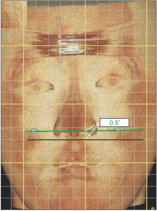
Fig. 7 Recreated natural head position (NHP) on three-dimensional (3D) simulator, frontal side. The 3D model was re-oriented to the NHP based on the roll and pitch data acquired by the device. Then the angle between two lingual buttons on both zygoma in the frontal side was calculated to compare the roll value.
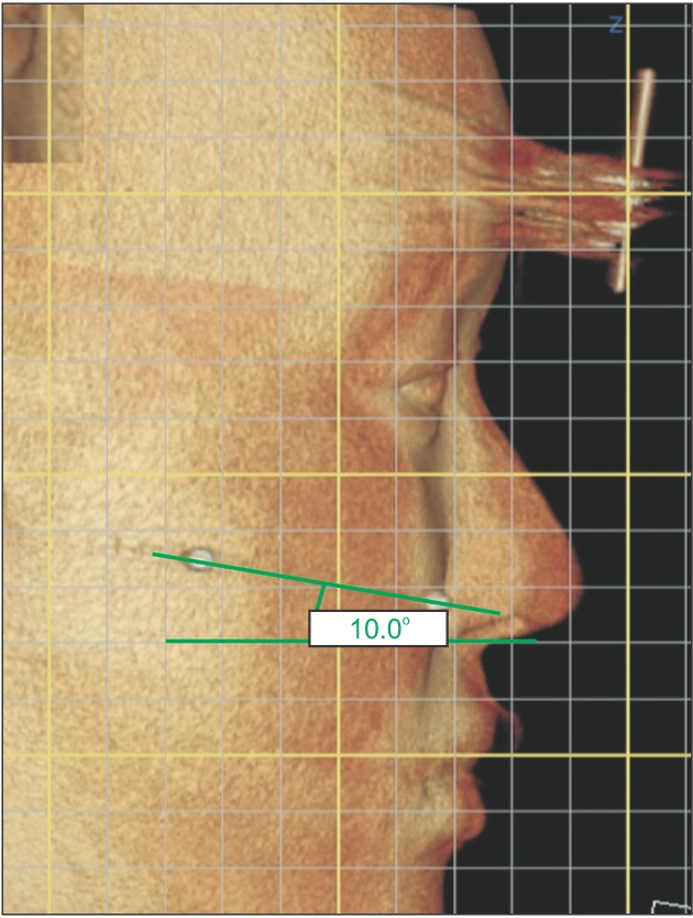
Fig. 8 Recreated natural head position (NHP) on three-dimensional (3D) simulator. The 3D model was re-oriented to the NHP based on the roll and pitch data acquired by the device. Then the angle between two lingual buttons on periauricula and zygomatic area in the lateral side was calculated to compare the pitch value.
Reference
-
1. Broca M. Sur les projections de la tète, et sur un nouveau procède de cephalometrié [The projections of the head and a new procedure in cephalometry]. Bull Soc Anthropol Paris. 1862; 3:514–544. French.2. Moorrees CFA, Kean MR. Natural head position, a basic consideration in the interpretation of cephalometric radiographs. Am J Phys Anthropol. 1958; 16:213–234.
Article3. Weber DW, Fallis DW, Packer MD. Three-dimensional reproducibility of natural head position. Am J Orthod Dentofacial Orthop. 2013; 143:738–744. PMID: 23631976.
Article4. Huggare JA, Laine-Alava MT. Nasorespiratory function and head posture. Am J Orthod Dentofacial Orthop. 1997; 112:507–511. PMID: 9387837.5. Lundström A. Natural head position/a discussion of concepts. Br J Orthod. 1990; 17:249–250. PMID: 2207058.6. Weber ZJ, Preston CB, Wright PG. Resistance to nasal airflow related to changes in head posture. Am J Orthod. 1981; 80:536–545. PMID: 6946709.
Article7. AlKofide EA, AlNamankani E. The association between posture of the head and malocclusion in Saudi subjects. Cranio. 2007; 25:98–105. PMID: 17508630.
Article8. Barbera AL, Sampson WJ, Townsend GC. Variation in natural head position and establishing corrected head position. Homo. 2014; 65:187–200. PMID: 24785580.
Article9. Andrighetto AR, de Fantini SM. Effects of neuromuscular deprogramming on the head position. Cranio. 2015; 33:183–188. PMID: 25052029.
Article10. Vig PS, Showfety KJ, Phillips C. Experimental manipulation of head posture. Am J Orthod. 1980; 77:258–268. PMID: 6928735.
Article11. Liu XJ, Li QQ, Pang YJ, Tian KY, Xie Z, Li ZL. Modified method of recording and reproducing natural head position with a multi-camera system and a laser level. Am J Orthod Dentofacial Orthop. 2015; 147:781–787. PMID: 26038082.
Article12. Schatz EC, Xia JJ, Gateno J, English JD, Teichgraeber JF, Garrett FA. Development of a technique for recording and transferring natural head position in 3 dimensions. J Craniofac Surg. 2010; 21:1452–1455. PMID: 20856035.
Article13. Lin XP, Arild S. Longitudinal study of the stability and reproducibility of natural head position in adolescents with different facial types over time. Shanghai Kou Qiang Yi Xue. 2005; 14:238–242. PMID: 15995767.14. Dubojska AM, Smiech-Slomkowska G. Natural head position and growth of the facial part of the skull. Cranio. 2013; 31:109–117. PMID: 23795400.
Article15. Showfety KJ, Vig PS, Matteson S. A simple method for taking natural-head-position cephalograms. Am J Orthod. 1983; 83:495–500. PMID: 6574706.
Article16. Viazis AD. A cephalometric analysis based on natural head position. J Clin Orthod. 1991; 25:172–181. PMID: 1939621.17. Leitão P, Nanda RS. Relationship of natural head position to craniofacial morphology. Am J Orthod Dentofacial Orthop. 2000; 117:406–417. PMID: 10756266.18. Robertson C. Cranial base considerations between apnoeics and non-apnoeic snorers, and associated effects of long-term mandibular advancement on condylar and natural head position. Eur J Orthod. 2002; 24:353–361. PMID: 12198865.
Article19. Uşümez S, Orhan M. Reproducibility of natural head position measured with an inclinometer. Am J Orthod Dentofacial Orthop. 2003; 123:451–454. PMID: 12695773.20. Cevidanes L, Oliveira AE, Motta A, Phillips C, Burke B, Tyndall D. Head orientation in CBCT-generated cephalograms. Angle Orthod. 2009; 79:971–977. PMID: 19705941.
Article21. Arnett GW, Gunson MJ. Facial planning for orthodontists and oral surgeons. Am J Orthod Dentofacial Orthop. 2004; 126:290–295. PMID: 15356488.
Article22. Uşümez S, Orhan M. Inclinometer method for recording and transferring natural head position in cephalometrics. Am J Orthod Dentofacial Orthop. 2001; 120:664–670. PMID: 11742312.23. Tian K, Li Q, Wang X, Liu X, Wang X, Li Z. Reproducibility of natural head position in normal Chinese people. Am J Orthod Dentofacial Orthop. 2015; 148:503–510. PMID: 26321348.
Article24. Swennen GR, Schutyser F, Barth EL, De Groeve P, De Mey A. A new method of 3-D cephalometry Part I: the anatomic Cartesian 3-D reference system. J Craniofac Surg. 2006; 17:314–325. PMID: 16633181.25. Xia JJ, McGrory JK, Gateno J, Teichgraeber JF, Dawson BC, Kennedy KA, et al. A new method to orient 3-dimensional computed tomography models to the natural head position: a clinical feasibility study. J Oral Maxillofac Surg. 2011; 69:584–591. PMID: 21353923.
Article26. Damstra J, Fourie Z, Ren Y. Simple technique to achieve a natural position of the head for cone beam computed tomography. Br J Oral Maxillofac Surg. 2010; 48:236–238. PMID: 19880225.
Article
- Full Text Links
-
- Actions
-
Cited
- CITED
-
- Close
- Share
-
- Similar articles
-
- The impact of reorienting cone-beam computed tomographic images in varied head positions on the coordinates of anatomical landmarks
- A study on the reproducibility of the natural head position according to the skeletal malocclusion type and sex
- Can Three-Dimensional Reconstructed Image Replace Medical Photograph?
- A cephalometric study of the natural head position according to craniofacial morphology
- The Supine and Prone Position for Computed Tomographic Myelography(CTM) of the Lumbar Spine: Change of Gantry Angle
