New method for an evaluation of the esthetical improvements resulting from a mandibular angle reduction
- Affiliations
-
- 1Department of Oral and Maxillofacial Surgery, College of Dentistry, Dankook University, Cheonan, Korea. hanimplant@dankook.ac.kr
- KMID: 2391354
- DOI: http://doi.org/10.5125/jkaoms.2017.43.4.239
Abstract
OBJECTIVES
This paper proposes Han's ratio as an objective and quantitative comparative result obtained from pre and postoperative data in patients with a mandibular angle reduction.
MATERIALS AND METHODS
Thirty patients, 12 men and 18 women, who visited the Department of Oral and Maxillofacial Surgery with the chief complaints of skeletal mandibular prognathism and prominent mandibular angle were selected. The subjects were classified into 3 groups according to the types of surgical procedures involved. Group A consisted of patients who underwent mandibular angle resection and mandibular setback. Group B was comprised of patients with mandibular angle resection, mandibular setback and genioplasty. Group C consisted of patients with mandibular angle resection, mandibular setback, Le Fort I osteotomy, and genioplasty. The landmarks placed in pre and postoperative frontal photographs were used to obtain the Han's ratio in each group. The Han's ratios were compared pre- and postoperation and according to the surgical techniques applied.
RESULTS
Of the 3 groups who had undergone a mandibular angle resection, all showed a statistically significant increase in Han's ratio. On the other hand, there was no statistically significant difference based on the surgical techniques used.
CONCLUSION
The ratio of the lateral lower face proposed in this study is a potential indicator of postoperative esthetic enhancement in mandibular angle reduction surgery.
MeSH Terms
Figure
-
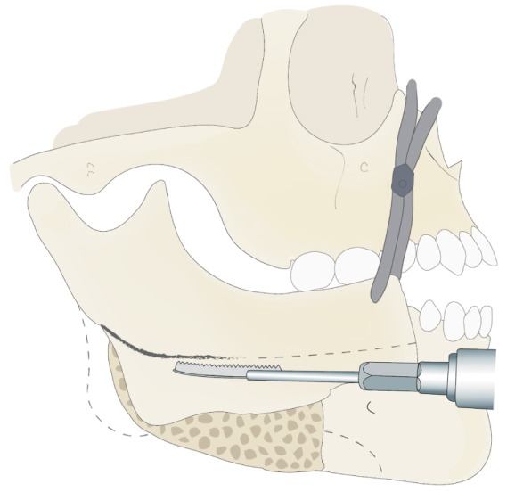
Fig. 1 Schematic drawing of curved ostectomy using reciprocating saw after sagittal split ramus osteotomy.
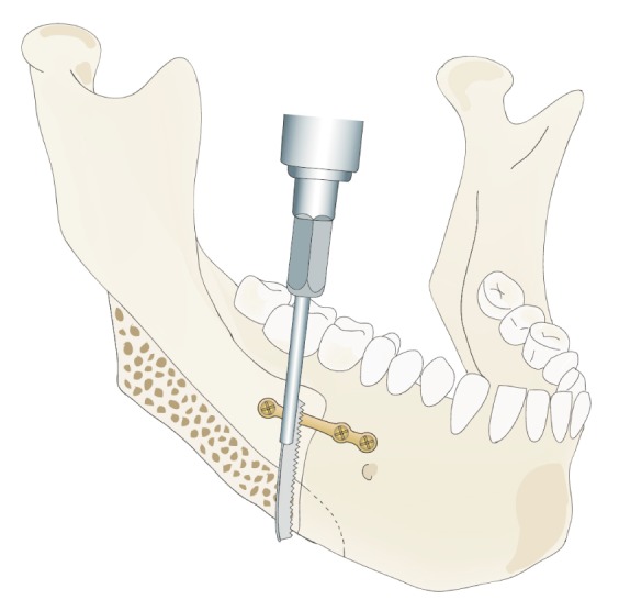
Fig. 2 Diagram of ostectomy design under the mental foramen after fixation of proximal segment to distal segment of mandible.
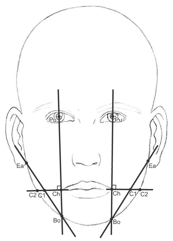
Fig. 3 Liner measurements and reference landmarks used in this study. (Pu: midpoint of pupil, Ch: most lateral point between upper and lower lip, C1: intersection of a line drawn from Ch perpendicular to Po-Bo and a line between Ba-Bo, C2: intersection of a line drawn from Ch perpendicular to Po-Bo and mandibular outline, Bo: mandibular outline, Ea: intersection of outlines of face and earlobe)
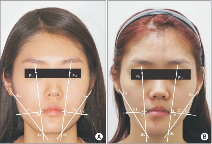
Fig. 4 Liner measurements and reference landmarks are showing in the preoperative (A) and postoperative (B) frontal facial photographs. Refer to Fig. 3 for the definition of landmarks.
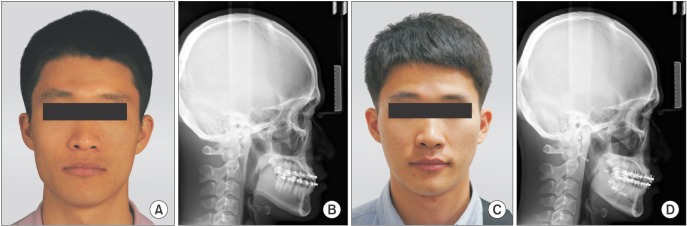
Fig. 5 Preoperative (A, B) and postoperative (C, D) changes of a frontal facial photograph and cephalometric lateral radiograph in Group A. The ratio increased from 0.67 to 0.72 after the surgery.
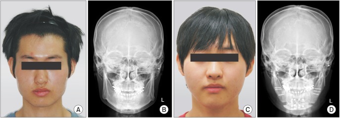
Fig. 6 Preoperative (A, B) and postoperative (C, D) changes of a frontal facial photograph and cephalometric posteroanterior radiograph in Group B. The ratio increased from 0.60 to 0.68 after the surgery.
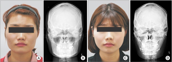
Fig. 7 Preoperative (A, B) and postoperative (C, D) changes of a frontal facial photograph and cephalometric posteroanterior radiograph in Group C. The ratio increased from 0.65 to 0.71 after the surgery.
Reference
-
1. Satoh K. Mandibular contouring surgery by angular contouring combined with genioplasty in orientals. Plast Reconstr Surg. 1998; 101:461–472. PMID: 9462783.
Article2. Coffey RJ. Unilateral hypertrophy of the masseter muscle. Surgery. 1942; 11:815.3. Rončević R. Masseter muscle hypertrophy: aetiology and therapy. J Oral Maxillofac Surg. 1986; 14:344–348.4. Goodwin DP, Calnan JS, McBride JA. Benign hypertrophy of the masseter muscles associated with hypofibrinogenaemia. A case report. Br J Plast Surg. 1967; 20:441–445. PMID: 4168494.5. Baek SM, Kim SS, Bindiger A. The prominent mandibular angle: preoperative management, operative technique, and results in 42 patients. Plast Reconstr Surg. 1989; 83:272–280. PMID: 2911627.6. Yang DB, Park CG. Mandibular contouring surgery for purely aesthetic reasons. Aesthetic Plast Surg. 1991; 15:53–60. PMID: 1994650.
Article7. Jin H. Misconceptions about mandible reduction procedures. Aesthetic Plast Surg. 2005; 29:317–324. PMID: 15959692.
Article8. Jin H, Kim BG. Mandibular angle reduction versus mandible reduction. Plast Reconstr Surg. 2004; 114:1263–1269. PMID: 15457047.
Article9. Han K, Kim J. Reduction mandibuloplasty: ostectomy of the lateral cortex around the mandibular angle. J Craniofac Surg. 2001; 12:314–325. PMID: 11482616.
Article10. Lee Y, Han K, Kang J. Korean standards of craniomaxillofacial skeleton. J Korean Soc Plast Reconstr Surg. 1994; 21:438–451.11. Choe KS, Sclafani AP, Litner JA, Yu GP, Romo T 3rd. The Korean American woman's face: anthropometric measurements and quantitative analysis of facial aesthetics. Arch Facial Plast Surg. 2004; 6:244–252. PMID: 15262719.12. Legg JW. Enlargement of the temporal and masseter muscle on both sides. Trans Pathol Soc. 1980; 31:361–366.13. Gurney CE. Chronic bilateral benign hypertrophy of the masseter muscles. Am J Surg. 1947; 73:137–139. PMID: 20279395.
Article14. Adams WM. Bilateral hypertrophy of the masseter muscle; an operation for correction; case report. Br J Plast Surg. 1949; 2:78–81. PMID: 18133192.15. Converse JM. Deformity of the jaws. In : Converse JM, editor. Reconstructive plastic surgery. Philadelphia: Saunders;1977. p. 1404.16. Baek SM, Baek RM, Shin MS. Refinement in aesthetic contouring of the prominent mandibular angle. Aesthetic Plast Surg. 1994; 18:283–289. PMID: 7976763.
Article17. Whitaker LA. Aesthetic contouring of the facial support system. Clin Plast Surg. 1989; 16:815–823. PMID: 2680223.
Article18. Cho JH, Han KH, Kang JS. Normal anthropometric values and standardized templates of Korean face and head. J Korean Soc Plast Reconstr Surg. 1993; 20:995–1005.19. Kim SK, Han JJ, Kim JT. Classification and treatment of prominent mandibular angle. Aesthetic Plast Surg. 2001; 25:382–387. PMID: 11692255.
Article
- Full Text Links
-
- Actions
-
Cited
- CITED
-
- Close
- Share
-
- Similar articles
-
- A study of post-operative changes in facial height and width of mandibular prognathic patients
- Reduction mandibular angleplasty assisted by c-arm fluoroscopy
- A Clinical Study of Mandibular Angle Fracture
- Mandibular angle reduction combined with facelift via the premasseter space
- The cephalometric study of korean mandibular angle
